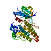
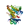
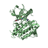
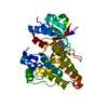
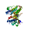
Entry Database : PDB / ID : 4pmpTitle The structure of TrkA kinase bound to the inhibitor 1-cyclopropyl-1-[3-(1,3-thiazol-2-yl)benzyl]-3-[4-(trifluoromethoxy)phenyl]urea High affinity nerve growth factor receptor Keywords / / Function / homology Function Domain/homology Component
/ / / / / / / / / / / / / / / / / / / / / / / / / / / / / / / / / / / / / / / / / / / / / / / / / / / / / / / / / / / / / / / / / / / / / / / / / / / / / / / / / / / / / / / / / / / / / / / / / / / / / / / / / / / / / / / / / / / / / / / / / / / / / / / / / / / / / / Biological species Homo sapiens (human)Method / / Resolution : 1.8 Å Authors Su, H.P. Journal : J.Med.Chem. / Year : 2014Title : Maximizing diversity from a kinase screen: identification of novel and selective pan-Trk inhibitors for chronic pain.Authors: Stachel, S.J. / Sanders, J.M. / Henze, D.A. / Rudd, M.T. / Su, H.P. / Li, Y. / Nanda, K.K. / Egbertson, M.S. / Manley, P.J. / Jones, K.L. / Brnardic, E.J. / Green, A. / Grobler, J.A. / ... Authors : Stachel, S.J. / Sanders, J.M. / Henze, D.A. / Rudd, M.T. / Su, H.P. / Li, Y. / Nanda, K.K. / Egbertson, M.S. / Manley, P.J. / Jones, K.L. / Brnardic, E.J. / Green, A. / Grobler, J.A. / Hanney, B. / Leitl, M. / Lai, M.T. / Munshi, V. / Murphy, D. / Rickert, K. / Riley, D. / Krasowska-Zoladek, A. / Daley, C. / Zuck, P. / Kane, S.A. / Bilodeau, M.T. History Deposition May 22, 2014 Deposition site / Processing site Revision 1.0 Jun 18, 2014 Provider / Type Revision 1.1 Oct 1, 2014 Group Revision 1.2 Feb 25, 2015 Group Revision 1.3 Dec 27, 2023 Group Data collection / Database references ... Data collection / Database references / Derived calculations / Other / Refinement description / Source and taxonomy Category chem_comp_atom / chem_comp_bond ... chem_comp_atom / chem_comp_bond / citation / database_2 / diffrn_radiation_wavelength / entity_src_gen / pdbx_database_status / pdbx_struct_assembly / pdbx_struct_oper_list / pdbx_struct_special_symmetry / refine_hist Item _citation.journal_id_CSD / _database_2.pdbx_DOI ... _citation.journal_id_CSD / _database_2.pdbx_DOI / _database_2.pdbx_database_accession / _entity_src_gen.pdbx_alt_source_flag / _pdbx_database_status.pdb_format_compatible / _pdbx_struct_assembly.oligomeric_details / _pdbx_struct_oper_list.symmetry_operation / _refine_hist.number_atoms_total / _refine_hist.pdbx_number_atoms_nucleic_acid / _refine_hist.pdbx_number_atoms_protein
Show all Show less
 Movie
Movie Controller

 Yorodumi
 Open data
 Basic information
 Components
 Keywords
 Function and homology information
 Homo sapiens (human)
 X-RAY DIFFRACTION /
  Authors
 Citation
 Structure visualization
 Downloads & links
 Other downloads
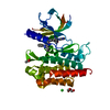


 PDBj
PDBj


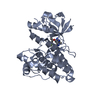
 Assembly
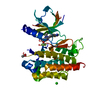

 Spodoptera frugiperda (fall armyworm)
 Mass: 433.447 Da / Num. of mol.: 1 / Source method: obtained synthetically / Formula: C21H18F3N3O2S
 Mass: 35.453 Da / Num. of mol.: 1 / Source method: obtained synthetically / Formula: Cl
 Mass: 59.044 Da / Num. of mol.: 3 / Source method: obtained synthetically / Formula: C2H3O2
 Mass: 18.015 Da / Num. of mol.: 171 / Source method: isolated from a natural source / Formula: H2O
 Sample preparation
 / Beamline: 17-ID / Wavelength: 1 Å
 Processing