[English] 日本語
Yorodumi
- PDB-4pl9: Structure of the catalytic domain of ETR1 from Arabidopsis thaliana -

+
Open data


ID or keywords:

Loading...

-
Basic information

Entry
Database: PDB / ID: 4pl9
TitleStructure of the catalytic domain of ETR1 from Arabidopsis thaliana
ComponentsEthylene receptor 1
KeywordsTRANSFERASE / ETR1 / Histidine Kinase / ethylene receptor / Cadmium / ADP
Function / homology
Function and homology information


ethylene receptor activity / regulation of seedling development / detection of ethylene stimulus / ethylene binding / seed dormancy process / defense response by callose deposition in cell wall / negative regulation of ethylene-activated signaling pathway / sugar mediated signaling pathway / response to gibberellin / response to insect ...ethylene receptor activity / regulation of seedling development / detection of ethylene stimulus / ethylene binding / seed dormancy process / defense response by callose deposition in cell wall / negative regulation of ethylene-activated signaling pathway / sugar mediated signaling pathway / response to gibberellin / response to insect / phloem or xylem histogenesis / cytokinin metabolic process / response to ethylene / regulation of stomatal movement / response to auxin / protein histidine kinase activity / response to abscisic acid / hydrogen peroxide biosynthetic process / histidine kinase / phosphorelay sensor kinase activity / response to salt stress / response to molecule of bacterial origin / defense response / response to heat / defense response to bacterium / cell division / endoplasmic reticulum membrane / endoplasmic reticulum / ATP binding / identical protein binding / metal ion binding
Similarity search - Function
Ethylene receptor / His Kinase A (phospho-acceptor) domain / His Kinase A (phosphoacceptor) domain / Signal transduction histidine kinase, dimerisation/phosphoacceptor domain / Signal transduction histidine kinase, dimerisation/phosphoacceptor domain superfamily / Signal transduction histidine kinase-related protein, C-terminal / Histidine kinase domain / Histidine kinase domain profile. / Response regulator receiver domain / cheY-homologous receiver domain ...Ethylene receptor / His Kinase A (phospho-acceptor) domain / His Kinase A (phosphoacceptor) domain / Signal transduction histidine kinase, dimerisation/phosphoacceptor domain / Signal transduction histidine kinase, dimerisation/phosphoacceptor domain superfamily / Signal transduction histidine kinase-related protein, C-terminal / Histidine kinase domain / Histidine kinase domain profile. / Response regulator receiver domain / cheY-homologous receiver domain / Histidine kinase-like ATPase, C-terminal domain / Signal transduction response regulator, receiver domain / Response regulatory domain profile. / Heat Shock Protein 90 / GAF domain / CheY-like superfamily / Domain present in phytochromes and cGMP-specific phosphodiesterases. / GAF domain / GAF-like domain superfamily / Histidine kinase-, DNA gyrase B-, and HSP90-like ATPase / Histidine kinase-like ATPases / Histidine kinase/HSP90-like ATPase / Histidine kinase/HSP90-like ATPase superfamily / 2-Layer Sandwich / Alpha Beta
Similarity search - Domain/homology
ACETATE ION / ADENOSINE-5'-DIPHOSPHATE / : / Ethylene receptor 1
Similarity search - Component
Biological speciesArabidopsis thaliana (thale cress)
MethodX-RAY DIFFRACTION / SYNCHROTRON / MOLECULAR REPLACEMENT / Resolution: 1.9 Å
AuthorsPanneerselvam, S. / Mueller-Dieckmann, J.
Citation
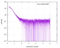
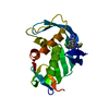
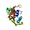
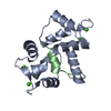
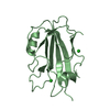
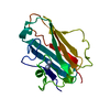
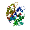
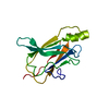
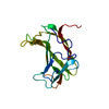
Journal: J Biol Chem / Year: 2015
Title: Structural model of the cytosolic domain of the plant ethylene receptor 1 (ETR1).
Authors: Hubert Mayerhofer / Saravanan Panneerselvam / Heidi Kaljunen / Anne Tuukkanen / Haydyn D T Mertens / Jochen Mueller-Dieckmann /
Abstract: Ethylene initiates important aspects of plant growth and development through disulfide-linked receptor dimers located in the endoplasmic reticulum. The receptors feature a small transmembrane, ...Ethylene initiates important aspects of plant growth and development through disulfide-linked receptor dimers located in the endoplasmic reticulum. The receptors feature a small transmembrane, ethylene binding domain followed by a large cytosolic domain, which serves as a scaffold for the assembly of large molecular weight complexes of different ethylene receptors and other cellular participants of the ethylene signaling pathway. Here we report the crystallographic structures of the ethylene receptor 1 (ETR1) catalytic ATP-binding and the ethylene response sensor 1 dimerization histidine phosphotransfer (DHp) domains and the solution structure of the entire cytosolic domain of ETR1, all from Arabidopsis thaliana. The isolated dimeric ethylene response sensor 1 DHp domain is asymmetric, the result of different helical bending angles close to the conserved His residue. The structures of the catalytic ATP-binding, DHp, and receiver domains of ethylene receptors and of a homologous, but dissimilar, GAF domain were refined against experimental small angle x-ray scattering data, leading to a structural model of the entire cytosolic domain of the ethylene receptor 1. The model illustrates that the cytosolic domain is shaped like a dumbbell and that the receiver domain is flexible and assumes a position different from those observed in prokaryotic histidine kinases. Furthermore the cytosolic domain of ETR1 plays a key role, interacting with all other receptors and several participants of the ethylene signaling pathway. Our model, therefore, provides the first step toward a detailed understanding of the molecular mechanics of this important signal transduction process in plants.
History
DepositionMay 16, 2014Deposition site: RCSB / Processing site: PDBE
Revision 1.0Dec 17, 2014Provider: repository / Type: Initial release
Revision 1.1Feb 11, 2015Group: Database references
Revision 1.2May 8, 2024Group: Data collection / Database references / Derived calculations
Category: chem_comp_atom / chem_comp_bond ...chem_comp_atom / chem_comp_bond / database_2 / pdbx_struct_conn_angle / pdbx_struct_special_symmetry / struct_conn
Item: _database_2.pdbx_DOI / _database_2.pdbx_database_accession ..._database_2.pdbx_DOI / _database_2.pdbx_database_accession / _pdbx_struct_conn_angle.ptnr1_auth_comp_id / _pdbx_struct_conn_angle.ptnr1_auth_seq_id / _pdbx_struct_conn_angle.ptnr1_label_asym_id / _pdbx_struct_conn_angle.ptnr1_label_atom_id / _pdbx_struct_conn_angle.ptnr1_label_comp_id / _pdbx_struct_conn_angle.ptnr1_label_seq_id / _pdbx_struct_conn_angle.ptnr2_symmetry / _pdbx_struct_conn_angle.ptnr3_auth_comp_id / _pdbx_struct_conn_angle.ptnr3_auth_seq_id / _pdbx_struct_conn_angle.ptnr3_label_asym_id / _pdbx_struct_conn_angle.ptnr3_label_atom_id / _pdbx_struct_conn_angle.ptnr3_label_comp_id / _pdbx_struct_conn_angle.ptnr3_label_seq_id / _pdbx_struct_conn_angle.value / _struct_conn.pdbx_dist_value / _struct_conn.pdbx_ptnr1_label_alt_id / _struct_conn.pdbx_ptnr2_label_alt_id / _struct_conn.ptnr1_auth_comp_id / _struct_conn.ptnr1_auth_seq_id / _struct_conn.ptnr1_label_asym_id / _struct_conn.ptnr1_label_atom_id / _struct_conn.ptnr1_label_comp_id / _struct_conn.ptnr1_label_seq_id / _struct_conn.ptnr2_auth_comp_id / _struct_conn.ptnr2_auth_seq_id / _struct_conn.ptnr2_label_asym_id / _struct_conn.ptnr2_label_atom_id / _struct_conn.ptnr2_label_comp_id / _struct_conn.ptnr2_symmetry

-
Structure visualization

Structure viewerMolecule:
MolmilJmol/JSmol

Downloads & links

-
Assembly

Deposited unit
A: Ethylene receptor 1
hetero molecules


Theoretical massNumber of molelcules
Total (without water)21,57015
Polymers19,8121
Non-polymers1,75814
Water1,78399
1


  • Idetical with deposited unit
  • defined by software
TypeNameSymmetry operationNumber
identity operation1_555x,y,z1
Buried area2130 Å2
ΔGint-73 kcal/mol
Surface area8320 Å2
MethodPISA
Unit cell
Length a, b, c (Å)76.260, 83.120, 91.930
Angle α, β, γ (deg.)90.00, 90.00, 90.00
Int Tables number24
Space group name H-MI212121
Components on special symmetry positions
IDModelComponents
11A-614-

ACT

21A-614-

ACT

31A-701-

HOH

41A-728-

HOH

-
Components

-
Protein , 1 types, 1 molecules A

#1: Protein Ethylene receptor 1 / AtETR1 / Protein ETHYLENE RESPONSE 1 / Protein ETR1


Mass: 19811.918 Da / Num. of mol.: 1 / Fragment: residue 407-589
Source method: isolated from a genetically manipulated source
Source: (gene. exp.) Arabidopsis thaliana (thale cress) / Gene: ETR1, At1g66340, T27F4.9 / Production host: Escherichia coli (E. coli) / References: UniProt: P49333, histidine kinase

-
Non-polymers , 5 types, 113 molecules

#2: Chemical
ChemComp-CD / CADMIUM ION


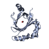
Mass: 112.411 Da / Num. of mol.: 11 / Source method: obtained synthetically / Formula: Cd
#3: Chemical ChemComp-CL / CHLORIDE ION


Mass: 35.453 Da / Num. of mol.: 1 / Source method: obtained synthetically / Formula: Cl
#4: Chemical ChemComp-ADP / ADENOSINE-5'-DIPHOSPHATE


Mass: 427.201 Da / Num. of mol.: 1 / Source method: obtained synthetically / Formula: C10H15N5O10P2 / Comment: ADP, energy-carrying molecule*YM
#5: Chemical ChemComp-ACT / ACETATE ION


Mass: 59.044 Da / Num. of mol.: 1 / Source method: obtained synthetically / Formula: C2H3O2
#6: Water ChemComp-HOH / water


Mass: 18.015 Da / Num. of mol.: 99 / Source method: isolated from a natural source / Formula: H2O

-
Experimental details

-
Experiment

ExperimentMethod: X-RAY DIFFRACTION

-
Sample preparation

CrystalDensity Matthews: 3.68 Å3/Da / Density % sol: 66.54 %
Crystal growTemperature: 292 K / Method: vapor diffusion, sitting drop / pH: 7.5
Details: 50 mM Cadmimum sulfate, 0.1 M HEPES pH 7.5, 1.0M Sodium acetate

-
Data collection

DiffractionMean temperature: 100 K
Diffraction sourceSource: SYNCHROTRON / Site: SLS / Beamline: X06DA / Wavelength: 0.998 Å
DetectorType: DECTRIS PILATUS 2M / Detector: PIXEL / Date: Feb 11, 2012
RadiationProtocol: SINGLE WAVELENGTH / Monochromatic (M) / Laue (L): M / Scattering type: x-ray
Radiation wavelengthWavelength: 0.998 Å / Relative weight: 1
ReflectionResolution: 1.9→42 Å / Num. obs: 23386 / % possible obs: 99.9 % / Redundancy: 5.6 % / Net I/σ(I): 26.3
Reflection shellResolution: 1.9→1.95 Å / Redundancy: 5.98 % / Mean I/σ(I) obs: 5 / % possible all: 100

-
Processing

Software
NameVersionClassification
PHENIX(phenix.refine: 1.8.1_1168)refinement
XDSdata reduction
SCALAdata scaling
SHELXDEphasing
RefinementMethod to determine structure: MOLECULAR REPLACEMENT / Resolution: 1.9→41.56 Å / SU ML: 0.14 / Cross valid method: FREE R-VALUE / σ(F): 1.36 / Phase error: 17.47 / Stereochemistry target values: ML
RfactorNum. reflection% reflectionSelection details
Rfree0.1816 1169 5 %Random selection
Rwork0.1465 ---
obs0.1483 23377 99.82 %-
Solvent computationShrinkage radii: 0.9 Å / VDW probe radii: 1.11 Å / Solvent model: FLAT BULK SOLVENT MODEL
Refinement stepCycle: LAST / Resolution: 1.9→41.56 Å
ProteinNucleic acidLigandSolventTotal
Num. atoms1154 0 43 99 1296
Refine LS restraints
Refine-IDTypeDev idealNumber
X-RAY DIFFRACTIONf_bond_d0.0171277
X-RAY DIFFRACTIONf_angle_d1.3191733
X-RAY DIFFRACTIONf_dihedral_angle_d16.243469
X-RAY DIFFRACTIONf_chiral_restr0.068203
X-RAY DIFFRACTIONf_plane_restr0.005208
LS refinement shell
Resolution (Å)Rfactor RfreeNum. reflection RfreeRfactor RworkNum. reflection RworkRefine-ID% reflection obs (%)
1.9-1.98640.21011440.13592748X-RAY DIFFRACTION100
1.9864-2.09120.1891450.12672762X-RAY DIFFRACTION100
2.0912-2.22220.15441430.12042715X-RAY DIFFRACTION100
2.2222-2.39370.1851450.12132754X-RAY DIFFRACTION100
2.3937-2.63460.19821450.13222755X-RAY DIFFRACTION100
2.6346-3.01570.20181470.15172788X-RAY DIFFRACTION100
3.0157-3.79910.18911470.15332790X-RAY DIFFRACTION100
3.7991-41.56990.16631530.15632896X-RAY DIFFRACTION99

+
About Yorodumi

-
News

-
Feb 9, 2022. New format data for meta-information of EMDB entries

New format data for meta-information of EMDB entries

  • Version 3 of the EMDB header file is now the official format.
  • The previous official version 1.9 will be removed from the archive.

Related info.:EMDB header

External links:wwPDB to switch to version 3 of the EMDB data model

-
Aug 12, 2020. Covid-19 info

Covid-19 info

URL: https://pdbjlvh1.pdbj.org/emnavi/covid19.php

New page: Covid-19 featured information page in EM Navigator.

Related info.:Covid-19 info / Mar 5, 2020. Novel coronavirus structure data

+
Mar 5, 2020. Novel coronavirus structure data

Novel coronavirus structure data

Related info.:Yorodumi Speices / Aug 12, 2020. Covid-19 info

External links:COVID-19 featured content - PDBj / Molecule of the Month (242):Coronavirus Proteases

+
Jan 31, 2019. EMDB accession codes are about to change! (news from PDBe EMDB page)

EMDB accession codes are about to change! (news from PDBe EMDB page)

  • The allocation of 4 digits for EMDB accession codes will soon come to an end. Whilst these codes will remain in use, new EMDB accession codes will include an additional digit and will expand incrementally as the available range of codes is exhausted. The current 4-digit format prefixed with “EMD-” (i.e. EMD-XXXX) will advance to a 5-digit format (i.e. EMD-XXXXX), and so on. It is currently estimated that the 4-digit codes will be depleted around Spring 2019, at which point the 5-digit format will come into force.
  • The EM Navigator/Yorodumi systems omit the EMD- prefix.

Related info.:Q: What is EMD? / ID/Accession-code notation in Yorodumi/EM Navigator

External links:EMDB Accession Codes are Changing Soon! / Contact to PDBj

+
Jul 12, 2017. Major update of PDB

Major update of PDB

  • wwPDB released updated PDB data conforming to the new PDBx/mmCIF dictionary.
  • This is a major update changing the version number from 4 to 5, and with Remediation, in which all the entries are updated.
  • In this update, many items about electron microscopy experimental information are reorganized (e.g. em_software).
  • Now, EM Navigator and Yorodumi are based on the updated data.

External links:wwPDB Remediation / Enriched Model Files Conforming to OneDep Data Standards Now Available in the PDB FTP Archive

-
Yorodumi

Thousand views of thousand structures

  • Yorodumi is a browser for structure data from EMDB, PDB, SASBDB, etc.
  • This page is also the successor to EM Navigator detail page, and also detail information page/front-end page for Omokage search.
  • The word "yorodu" (or yorozu) is an old Japanese word meaning "ten thousand". "mi" (miru) is to see.

Related info.:EMDB / PDB / SASBDB / Comparison of 3 databanks / Yorodumi Search / Aug 31, 2016. New EM Navigator & Yorodumi / Yorodumi Papers / Jmol/JSmol / Function and homology information / Changes in new EM Navigator and Yorodumi

Read more