 ムービー
ムービー コントローラー


+ データを開く

- 基本情報
基本情報
| 登録情報 | データベース: PDB / ID: 4pb6 | ||||||
|---|---|---|---|---|---|---|---|
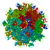
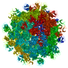
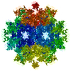
| タイトル | Feline calicivirus VP1 T=1 virus-like particle | ||||||
 要素 要素 | VP1 | ||||||
 キーワード キーワード | VIRUS / viral capsid protein / virus-like particle | ||||||
| 機能・相同性 | T=3 icosahedral viral capsid / Calicivirus coat protein / Calicivirus coat protein / Picornavirus/Calicivirus coat protein / Viral coat protein subunit / host cell cytoplasm / Capsid protein VP1 機能・相同性情報 機能・相同性情報 | ||||||
| 生物種 |  Feline calicivirus (ウイルス) Feline calicivirus (ウイルス) | ||||||
| 手法 |  X線回折 / シンクロトロン / 分子置換 / 解像度: 8 Å X線回折 / シンクロトロン / 分子置換 / 解像度: 8 Å | ||||||
 データ登録者 データ登録者 | Burmeister, W.P. / Buisson, M. | ||||||
 引用 引用 | ジャーナル: PLoS One / 年: 2015 タイトル: Structure determination of feline calicivirus virus-like particles in the context of a pseudo-octahedral arrangement. 著者: Wim P Burmeister / Marlyse Buisson / Leandro F Estrozi / Guy Schoehn / Olivier Billet / Zahia Hannas / Cécile Sigoillot / Hervé Poulet /  要旨: The vesivirus feline calicivirus (FCV) is a positive strand RNA virus encapsidated by an icosahedral T=3 shell formed by the viral VP1 protein. Upon its expression in the insect cell - baculovirus ...The vesivirus feline calicivirus (FCV) is a positive strand RNA virus encapsidated by an icosahedral T=3 shell formed by the viral VP1 protein. Upon its expression in the insect cell - baculovirus system in the context of vaccine development, two types of virus-like particles (VLPs) were formed, a majority built of 60 subunits (T=1) and a minority probably built of 180 subunits (T=3). The structure of the small particles was determined by x-ray crystallography at 0.8 nm resolution helped by cryo-electron microscopy in order to understand their formation. Cubic crystals belonged to space group P213. Their self-rotation function showed the presence of an octahedral pseudo-symmetry similar to the one described previously by Agerbandje and co-workers for human parvovirus VLPs. The crystal structure could be solved starting from the published VP1 structure in the context of the T=3 viral capsid. In contrast to viral capsids, where the capsomers are interlocked by the exchange of the N-terminal arm (NTA) domain, this domain is disordered in the T=1 capsid of the VLPs. Furthermore it is prone to proteolytic cleavage. The relative orientation of P (protrusion) and S (shell) domains is alerted so as to fit VP1 to the smaller T=1 particle whereas the intermolecular contacts around 2-fold, 3-fold and 5-fold axes are conserved. By consequence the surface of the VLP is very similar compared to the viral capsid and suggests a similar antigenicity. The knowledge of the structure of the VLPs will help to improve their stability, in respect to a use for vaccination. | ||||||
| 履歴 |
|
- 構造の表示
構造の表示
| 構造ビューア | 分子: MolmilJmol/JSmol |
|---|
- ダウンロードとリンク
-ダウンロード
| PDBx/mmCIF形式 | 4pb6.cif.gz | 1.6 MB | 表示 | PDBx/mmCIF形式 |
|---|---|---|---|---|
| PDB形式 | pdb4pb6.ent.gz | 1.3 MB | 表示 | PDB形式 |
| PDBx/mmJSON形式 | 4pb6.json.gz | ツリー表示 | PDBx/mmJSON形式 | |
| その他 |  その他のダウンロード その他のダウンロード |
-検証レポート
| 文書・要旨 | 4pb6_validation.pdf.gz | 737.8 KB | 表示 | wwPDB検証レポート |
|---|---|---|---|---|
| 文書・詳細版 | 4pb6_full_validation.pdf.gz | 2.1 MB | 表示 | |
| XML形式データ | 4pb6_validation.xml.gz | 518.9 KB | 表示 | |
| CIF形式データ | 4pb6_validation.cif.gz | 669.3 KB | 表示 | |
| アーカイブディレクトリ | https://data.pdbj.org/pub/pdb/validation_reports/pb/4pb6ftp://data.pdbj.org/pub/pdb/validation_reports/pb/4pb6 | HTTPS FTP |
-関連構造データ


-リンク
 PDBj
PDBj

- 集合体
集合体
| 登録構造単位 | 
| ||||||||||||
|---|---|---|---|---|---|---|---|---|---|---|---|---|---|
| 1 | 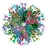
| ||||||||||||
| 2 |
| ||||||||||||
| 3 | 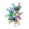
| ||||||||||||
| 単位格子 |
| ||||||||||||
| 対称性 | 点対称性: (シェーンフリース記号: C3 (3回回転対称)) | ||||||||||||
| 非結晶学的対称性 (NCS) | NCS oper:
|
-要素
| #1: タンパク質 | 分子量: 59297.613 Da / 分子数: 20 / 由来タイプ: 組換発現 / 由来: (組換発現) Feline calicivirus (ウイルス) / 株: Merial100869 / 遺伝子: VP1 / 細胞株 (発現宿主): Sf9発現宿主:   Spodoptera frugiperda (ツマジロクサヨトウ) Spodoptera frugiperda (ツマジロクサヨトウ)参照: UniProt: A0A0J9X1Z3*PLUS |
|---|
-実験情報
-実験
| 実験 | 手法: X線回折 |
|---|
- 試料調製
試料調製
| 結晶 | マシュー密度: 3.24 Å3/Da / 溶媒含有率: 62 % / 解説: tetrahedra or octahedra |
|---|---|
| 結晶化 | 温度: 293 K / 手法: 蒸気拡散法, ハンギングドロップ法 / pH: 7 詳細: reservoir solution: 7 - 10 % PEG 6000, 0.1 M Hepes pH 7, 0.5 - 1 M LiCl protein in: 20 mM MES pH 6, 200 mM NaCl at 3.4 mg/ml |
-データ収集
| 回折 | 平均測定温度: 100 K |
|---|---|
| 放射光源 | 由来: シンクロトロン / サイト: ESRF / ビームライン: ID23-2 / 波長: 0.9185 Å |
| 検出器 | タイプ: ADSC QUANTUM 4 / 検出器: CCD / 日付: 2010年2月15日 |
| 放射 | プロトコル: SINGLE WAVELENGTH / 単色(M)・ラウエ(L): M / 散乱光タイプ: x-ray |
| 放射波長 | 波長: 0.9185 Å / 相対比: 1 |
| 反射 | 解像度: 8→67.2 Å / Num. all: 14809 / Num. obs: 14809 / % possible obs: 87.22 % / 冗長度: 5.5 % / Rmerge(I) obs: 0.24 / Rsym value: 0.24 / Net I/av σ(I): 6.64 / Net I/σ(I): 3.07 |
| 反射 シェル | 解像度: 8→8.43 Å / 冗長度: 1.82 % / Rmerge(I) obs: 1.1 / Mean I/σ(I) obs: 0.69 / % possible all: 61.5 |
- 解析
解析
| ソフトウェア |
| |||||||||||||||||||||
|---|---|---|---|---|---|---|---|---|---|---|---|---|---|---|---|---|---|---|---|---|---|---|
| 精密化 | 構造決定の手法: 分子置換 開始モデル: pdb 3m8l and model builting 解像度: 8→65.51 Å / Cor.coef. Fo:Fc: 0.503 / Cor.coef. Fo:Fc free: 0.42 / SU B: 0.003 / SU ML: 0 / 交差検証法: THROUGHOUT / ESU R: 8.911 / ESU R Free: 9.346 / 立体化学のターゲット値: MAXIMUM LIKELIHOOD / 詳細: HYDROGENS HAVE BEEN USED IF PRESENT IN THE INPUT
| |||||||||||||||||||||
| 溶媒の処理 | イオンプローブ半径: 0.8 Å / 減衰半径: 0.8 Å / VDWプローブ半径: 1.2 Å / 溶媒モデル: BABINET MODEL WITH MASK | |||||||||||||||||||||
| 原子変位パラメータ | Biso mean: 94.995 Å2
| |||||||||||||||||||||
| 精密化ステップ | サイクル: 1 / 解像度: 8→65.51 Å
| |||||||||||||||||||||
| LS精密化 シェル | Refine-ID: X-RAY DIFFRACTION
|
