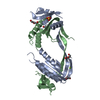
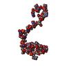
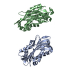
Entry Database : PDB / ID : 4p2mTitle Swapped Dimer of Mycobacterial Adenylyl cyclase Rv1625c: Form 1 Adenylate cyclase Keywords / / / / Function / homology Function Domain/homology Component
/ / / / / / / / / / / / / / / / / / / / / / / / / / / / / / / / / / / / / / / / / / / / Biological species Mycobacterium tuberculosis (bacteria)Method / / / Resolution : 2.7 Å Authors Barathy, D.V. / Mattoo, R. / Visweswariah, S.S. / Suguna, K. Journal : Iucrj / Year : 2014Title : New structural forms of a mycobacterial adenylyl cyclase Rv1625c.Authors : Barathy, D. / Mattoo, R. / Visweswariah, S. / Suguna, K. History Deposition Mar 4, 2014 Deposition site / Processing site Revision 1.0 Sep 17, 2014 Provider / Type Revision 1.1 Oct 22, 2014 Group Revision 1.2 Nov 1, 2017 Group Author supporting evidence / Derived calculations ... Author supporting evidence / Derived calculations / Other / Refinement description / Source and taxonomy / Structure summary Category entity_src_gen / pdbx_database_status ... entity_src_gen / pdbx_database_status / pdbx_struct_assembly / pdbx_struct_assembly_auth_evidence / pdbx_struct_oper_list / software / struct_keywords Item _entity_src_gen.pdbx_alt_source_flag / _pdbx_database_status.pdb_format_compatible ... _entity_src_gen.pdbx_alt_source_flag / _pdbx_database_status.pdb_format_compatible / _pdbx_struct_assembly.oligomeric_details / _pdbx_struct_oper_list.symmetry_operation / _software.classification / _struct_keywords.text Revision 1.3 Sep 27, 2023 Group / Database references / Refinement descriptionCategory chem_comp_atom / chem_comp_bond ... chem_comp_atom / chem_comp_bond / database_2 / pdbx_initial_refinement_model / refine_hist Item _database_2.pdbx_DOI / _database_2.pdbx_database_accession ... _database_2.pdbx_DOI / _database_2.pdbx_database_accession / _refine_hist.number_atoms_solvent / _refine_hist.pdbx_number_atoms_ligand / _refine_hist.pdbx_number_atoms_nucleic_acid / _refine_hist.pdbx_number_atoms_protein
Show all Show less
 Movie
Movie Controller


 Open data
 Basic information
 Components
 Keywords
 Function and homology information
 Mycobacterium tuberculosis (bacteria)
 X-RAY DIFFRACTION /
  Authors
 Citation
 Structure visualization
 Downloads & links
 Other downloads
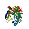


 PDBj
PDBj


 Assembly


 Mass: 96.063 Da / Num. of mol.: 2 / Source method: obtained synthetically / Formula: SO4
 Mass: 106.120 Da / Num. of mol.: 9 / Source method: obtained synthetically / Formula: C4H10O3
 Mass: 18.015 Da / Num. of mol.: 65 / Source method: isolated from a natural source / Formula: H2O
 Sample preparation
 / Beamline: BM14 / Wavelength: 0.9762 Å
 Processing