 Movie
Movie Controller

[English] 日本語
 Yorodumi
- PDB-4ozr: Crystal structure of the ligand binding domains of the Bovicola o... -
+ Open data

- Basic information
| Entry | Database: PDB / ID: 4ozr | ||||||
|---|---|---|---|---|---|---|---|
| Title | Crystal structure of the ligand binding domains of the Bovicola ovis ecdysone receptor EcR/USP heterodimer (methylene lactam crystal) | ||||||
 Components Components |
| ||||||
 Keywords Keywords | TRANSCRIPTION / Ecdysone receptor / USP / methylene lactam / heterodimer / ligand binding domain | ||||||
| Function / homology |  Function and homology information Function and homology informationecdysone binding / ecdysone receptor signaling pathway / nuclear steroid receptor activity / nuclear receptor activity / sequence-specific DNA binding / DNA-binding transcription factor activity / zinc ion binding / nucleus Similarity search - Function | ||||||
| Biological species |  Pediculus humanus subsp. corporis (human body louse) Pediculus humanus subsp. corporis (human body louse) | ||||||
| Method |  X-RAY DIFFRACTION / SYNCHROTRON / MOLECULAR REPLACEMENT / Resolution: 2.7 Å X-RAY DIFFRACTION / SYNCHROTRON / MOLECULAR REPLACEMENT / Resolution: 2.7 Å | ||||||
 Authors Authors | Ren, B. / Peat, T.S. / Streltsov, V.A. / Pollard, M. / Fernley, R. / Grusovin, J. / Seabrook, S. / Pilling, P. / Phan, T. / Lu, L. ...Ren, B. / Peat, T.S. / Streltsov, V.A. / Pollard, M. / Fernley, R. / Grusovin, J. / Seabrook, S. / Pilling, P. / Phan, T. / Lu, L. / Lovrecz, G.O. / Graham, L.D. / Hill, R.J. | ||||||
 Citation Citation | Journal: Acta Crystallogr.,Sect.D / Year: 2014 Title: Unprecedented conformational flexibility revealed in the ligand-binding domains of the Bovicola ovis ecdysone receptor (EcR) and ultraspiracle (USP) subunits. Authors: Ren, B. / Peat, T.S. / Streltsov, V.A. / Pollard, M. / Fernley, R. / Grusovin, J. / Seabrook, S. / Pilling, P. / Phan, T. / Lu, L. / Lovrecz, G.O. / Graham, L.D. / Hill, R.J. | ||||||
| History |
|
- Structure visualization
| Structure viewer | Molecule: MolmilJmol/JSmol |
|---|
- Downloads & links
-Download
| PDBx/mmCIF format | 4ozr.cif.gz | 175.3 KB | Display | PDBx/mmCIF format |
|---|---|---|---|---|
| PDB format | pdb4ozr.ent.gz | 138.6 KB | Display | PDB format |
| PDBx/mmJSON format | 4ozr.json.gz | Tree view | PDBx/mmJSON format | |
| Others |  Other downloads Other downloads |
-Validation report
| Summary document | 4ozr_validation.pdf.gz | 437.7 KB | Display | wwPDB validaton report |
|---|---|---|---|---|
| Full document | 4ozr_full_validation.pdf.gz | 447.3 KB | Display | |
| Data in XML | 4ozr_validation.xml.gz | 17.3 KB | Display | |
| Data in CIF | 4ozr_validation.cif.gz | 23.5 KB | Display | |
| Arichive directory | https://data.pdbj.org/pub/pdb/validation_reports/oz/4ozrftp://data.pdbj.org/pub/pdb/validation_reports/oz/4ozr | HTTPS FTP |
-Related structure data

-Links
 PDBj
PDBj
- Assembly
| Deposited unit | 
| ||||||||
|---|---|---|---|---|---|---|---|---|---|
| 1 |
| ||||||||
| 2 | 
| ||||||||
| 3 | 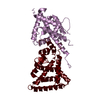
| ||||||||
| Unit cell |
|
-Components
| #1: Protein | Mass: 22935.762 Da / Num. of mol.: 1 / Fragment: UNP residues 281-514 Source method: isolated from a genetically manipulated source Source: (gene. exp.) Pediculus humanus subsp. corporis (human body louse)Gene: Phum_PHUM467460 / Production host:  |
|---|---|
| #2: Protein | Mass: 22074.666 Da / Num. of mol.: 1 / Fragment: UNP residues 222-415 Source method: isolated from a genetically manipulated source Source: (gene. exp.) Pediculus humanus subsp. corporis (human body louse)Gene: Phum_PHUM164330 / Production host: |
| #3: Water | ChemComp-HOH /  Mass: 18.015 Da / Num. of mol.: 91 / Source method: isolated from a natural source / Formula: H2O Mass: 18.015 Da / Num. of mol.: 91 / Source method: isolated from a natural source / Formula: H2O |
-Experimental details
-Experiment
| Experiment | Method: X-RAY DIFFRACTION |
|---|
- Sample preparation
| Crystal | Density Matthews: 2.78 Å3/Da / Density % sol: 55.74 % |
|---|---|
| Crystal grow | Temperature: 293 K / Method: vapor diffusion, sitting drop Details: 50 mM Tris pH 7.5, 150 mM NaCl, 5% glycerol, 5 mM DTT, 0.02% azide, 0.37 mM methylene lactam PH range: 7.5 |
-Data collection
| Diffraction | Mean temperature: 100 K |
|---|---|
| Diffraction source | Source: SYNCHROTRON / Site: Australian Synchrotron  / Beamline: MX2 / Wavelength: 0.9536 Å / Beamline: MX2 / Wavelength: 0.9536 Å |
| Detector | Type: ADSC QUANTUM 315r / Detector: CCD / Date: Oct 21, 2011 |
| Radiation | Protocol: SINGLE WAVELENGTH / Scattering type: x-ray |
| Radiation wavelength | Wavelength: 0.9536 Å / Relative weight: 1 |
| Reflection | Resolution: 2.7→69.3 Å / Num. obs: 13311 / % possible obs: 96.2 % / Redundancy: 2.5 % / Net I/σ(I): 9.1 |
- Processing
| Software | Name: PHENIX / Version: (phenix.refine: 1.8.1_1168) / Classification: refinement | |||||||||||||||||||||||||||||||||||||||||||||||||||||||||||||||||||||||||||||||||||||||||||||||||||||||||||||||||||||||||||||||||||||||||||||||||||||||||||||||||||||||||||||||||||||||||||||||||||||||||||||||||||||||||||||||||||||||||||||||||||||||||||||||||||||||||||||||||||||||||||||||||||||||||||||||||||||||||||||||||||||
|---|---|---|---|---|---|---|---|---|---|---|---|---|---|---|---|---|---|---|---|---|---|---|---|---|---|---|---|---|---|---|---|---|---|---|---|---|---|---|---|---|---|---|---|---|---|---|---|---|---|---|---|---|---|---|---|---|---|---|---|---|---|---|---|---|---|---|---|---|---|---|---|---|---|---|---|---|---|---|---|---|---|---|---|---|---|---|---|---|---|---|---|---|---|---|---|---|---|---|---|---|---|---|---|---|---|---|---|---|---|---|---|---|---|---|---|---|---|---|---|---|---|---|---|---|---|---|---|---|---|---|---|---|---|---|---|---|---|---|---|---|---|---|---|---|---|---|---|---|---|---|---|---|---|---|---|---|---|---|---|---|---|---|---|---|---|---|---|---|---|---|---|---|---|---|---|---|---|---|---|---|---|---|---|---|---|---|---|---|---|---|---|---|---|---|---|---|---|---|---|---|---|---|---|---|---|---|---|---|---|---|---|---|---|---|---|---|---|---|---|---|---|---|---|---|---|---|---|---|---|---|---|---|---|---|---|---|---|---|---|---|---|---|---|---|---|---|---|---|---|---|---|---|---|---|---|---|---|---|---|---|---|---|---|---|---|---|---|---|---|---|---|---|---|---|---|---|---|---|---|---|---|---|---|---|---|---|---|---|---|---|---|---|---|---|---|---|---|---|---|---|---|---|---|---|---|---|---|---|---|---|---|---|---|---|---|---|---|---|---|---|---|---|---|---|---|---|
| Refinement | Method to determine structure: MOLECULAR REPLACEMENT / Resolution: 2.7→68.03 Å / SU ML: 0.37 / Cross valid method: FREE R-VALUE / σ(F): 1.34 / Phase error: 27.91 / Stereochemistry target values: ML
| |||||||||||||||||||||||||||||||||||||||||||||||||||||||||||||||||||||||||||||||||||||||||||||||||||||||||||||||||||||||||||||||||||||||||||||||||||||||||||||||||||||||||||||||||||||||||||||||||||||||||||||||||||||||||||||||||||||||||||||||||||||||||||||||||||||||||||||||||||||||||||||||||||||||||||||||||||||||||||||||||||||
| Solvent computation | Shrinkage radii: 0.7 Å / VDW probe radii: 1.1 Å / Solvent model: FLAT BULK SOLVENT MODEL | |||||||||||||||||||||||||||||||||||||||||||||||||||||||||||||||||||||||||||||||||||||||||||||||||||||||||||||||||||||||||||||||||||||||||||||||||||||||||||||||||||||||||||||||||||||||||||||||||||||||||||||||||||||||||||||||||||||||||||||||||||||||||||||||||||||||||||||||||||||||||||||||||||||||||||||||||||||||||||||||||||||
| Refinement step | Cycle: LAST / Resolution: 2.7→68.03 Å
| |||||||||||||||||||||||||||||||||||||||||||||||||||||||||||||||||||||||||||||||||||||||||||||||||||||||||||||||||||||||||||||||||||||||||||||||||||||||||||||||||||||||||||||||||||||||||||||||||||||||||||||||||||||||||||||||||||||||||||||||||||||||||||||||||||||||||||||||||||||||||||||||||||||||||||||||||||||||||||||||||||||
| Refine LS restraints |
| |||||||||||||||||||||||||||||||||||||||||||||||||||||||||||||||||||||||||||||||||||||||||||||||||||||||||||||||||||||||||||||||||||||||||||||||||||||||||||||||||||||||||||||||||||||||||||||||||||||||||||||||||||||||||||||||||||||||||||||||||||||||||||||||||||||||||||||||||||||||||||||||||||||||||||||||||||||||||||||||||||||
| LS refinement shell |
| |||||||||||||||||||||||||||||||||||||||||||||||||||||||||||||||||||||||||||||||||||||||||||||||||||||||||||||||||||||||||||||||||||||||||||||||||||||||||||||||||||||||||||||||||||||||||||||||||||||||||||||||||||||||||||||||||||||||||||||||||||||||||||||||||||||||||||||||||||||||||||||||||||||||||||||||||||||||||||||||||||||
| Refinement TLS params. | Method: refined / Refine-ID: X-RAY DIFFRACTION
| |||||||||||||||||||||||||||||||||||||||||||||||||||||||||||||||||||||||||||||||||||||||||||||||||||||||||||||||||||||||||||||||||||||||||||||||||||||||||||||||||||||||||||||||||||||||||||||||||||||||||||||||||||||||||||||||||||||||||||||||||||||||||||||||||||||||||||||||||||||||||||||||||||||||||||||||||||||||||||||||||||||
| Refinement TLS group |
|
