 Movie
Movie Controller

[English] 日本語
 Yorodumi
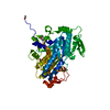
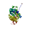
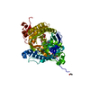
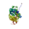
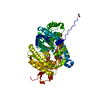
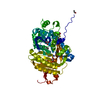
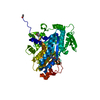
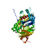
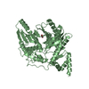
- PDB-4oyi: Human solAC Complexed with (4-Amino-furazan-3-yl)-phenyl-methanone -
+ Open data

- Basic information
| Entry | Database: PDB / ID: 4oyi | ||||||
|---|---|---|---|---|---|---|---|
| Title | Human solAC Complexed with (4-Amino-furazan-3-yl)-phenyl-methanone | ||||||
 Components Components | Adenylate cyclase type 10 | ||||||
 Keywords Keywords | LYASE | ||||||
| Function / homology |  Function and homology information Function and homology informationnegative regulation of cardiac muscle cell contraction / astrocyte end-foot / mitochondrial ATP transmembrane transport / bicarbonate binding / epithelial cilium movement involved in extracellular fluid movement / neuron projection retraction / positive regulation of glycogen catabolic process / : / central region of growth cone / glucose catabolic process ...negative regulation of cardiac muscle cell contraction / astrocyte end-foot / mitochondrial ATP transmembrane transport / bicarbonate binding / epithelial cilium movement involved in extracellular fluid movement / neuron projection retraction / positive regulation of glycogen catabolic process / : / central region of growth cone / glucose catabolic process / regulation of mitophagy / regulation of membrane repolarization / adenylate cyclase / basal part of cell / positive regulation of oxidative stress-induced neuron intrinsic apoptotic signaling pathway / cAMP biosynthetic process / positive regulation of ossification / adenylate cyclase activity / positive regulation of protein targeting to mitochondrion / positive regulation of reactive oxygen species biosynthetic process / neuron projection extension / positive regulation of vascular associated smooth muscle cell apoptotic process / positive regulation of mitochondrial depolarization / positive regulation of ATP biosynthetic process / positive regulation of cardiac muscle hypertrophy / positive regulation of cardiac muscle cell apoptotic process / negative regulation of mitochondrial membrane potential / spermatid development / positive regulation of axon extension / Hedgehog 'off' state / negative regulation of reactive oxygen species biosynthetic process / neuron projection maintenance / positive regulation of peptidyl-threonine phosphorylation / cilium / manganese ion binding / ATPase binding / cytoskeleton / intracellular signal transduction / apical plasma membrane / neuronal cell body / dendrite / perinuclear region of cytoplasm / magnesium ion binding / mitochondrion / extracellular region / ATP binding / nucleus / cytoplasm / cytosol Similarity search - Function | ||||||
| Biological species |  Homo sapiens (human) Homo sapiens (human) | ||||||
| Method |  X-RAY DIFFRACTION / SYNCHROTRON / Resolution: 1.7 Å X-RAY DIFFRACTION / SYNCHROTRON / Resolution: 1.7 Å | ||||||
 Authors Authors | Vinkovic, M. | ||||||
 Citation Citation | Journal: Chemmedchem / Year: 2014 Title: Crystal structure of human soluble adenylate cyclase reveals a distinct, highly flexible allosteric bicarbonate binding pocket. Authors: Saalau-Bethell, S.M. / Berdini, V. / Cleasby, A. / Congreve, M. / Coyle, J.E. / Lock, V. / Murray, C.W. / O'Brien, M.A. / Rich, S.J. / Sambrook, T. / Vinkovic, M. / Yon, J.R. / Jhoti, H. | ||||||
| History |
|
- Structure visualization
| Structure viewer | Molecule: MolmilJmol/JSmol |
|---|
- Downloads & links
-Download
| PDBx/mmCIF format | 4oyi.cif.gz | 213.7 KB | Display | PDBx/mmCIF format |
|---|---|---|---|---|
| PDB format | pdb4oyi.ent.gz | 168.8 KB | Display | PDB format |
| PDBx/mmJSON format | 4oyi.json.gz | Tree view | PDBx/mmJSON format | |
| Others |  Other downloads Other downloads |
-Validation report
| Summary document | 4oyi_validation.pdf.gz | 457 KB | Display | wwPDB validaton report |
|---|---|---|---|---|
| Full document | 4oyi_full_validation.pdf.gz | 469.6 KB | Display | |
| Data in XML | 4oyi_validation.xml.gz | 27.5 KB | Display | |
| Data in CIF | 4oyi_validation.cif.gz | 42.2 KB | Display | |
| Arichive directory | https://data.pdbj.org/pub/pdb/validation_reports/oy/4oyiftp://data.pdbj.org/pub/pdb/validation_reports/oy/4oyi | HTTPS FTP |
-Related structure data
| Related structure data |  4oyaC  4oybC  4oymC  4oyoC  4oypC  4oywC  4oyxC  4oyzC  4oz2C  4oz3C C: citing same article ( |
|---|---|
| Similar structure data |

-Links
 PDBj
PDBj


- Assembly
| Deposited unit | 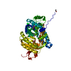
| |||||||||
|---|---|---|---|---|---|---|---|---|---|---|
| 1 |
| |||||||||
| 2 | 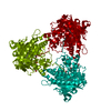
| |||||||||
| Unit cell |
| |||||||||
| Components on special symmetry positions |
|
-Components
| #1: Protein | Mass: 53466.832 Da / Num. of mol.: 1 Source method: isolated from a genetically manipulated source Source: (gene. exp.) Homo sapiens (human) / Gene: ADCY10, SAC / Production host:   Spodoptera frugiperda (fall armyworm) / References: UniProt: Q96PN6, adenylate cyclase Spodoptera frugiperda (fall armyworm) / References: UniProt: Q96PN6, adenylate cyclase | ||
|---|---|---|---|
| #2: Chemical | ChemComp-1VK / (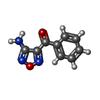  Mass: 189.171 Da / Num. of mol.: 1 / Source method: obtained synthetically / Formula: C9H7N3O2 Mass: 189.171 Da / Num. of mol.: 1 / Source method: obtained synthetically / Formula: C9H7N3O2 | ||
| #3: Chemical |   Mass: 92.094 Da / Num. of mol.: 2 / Source method: obtained synthetically / Formula: C3H8O3 Mass: 92.094 Da / Num. of mol.: 2 / Source method: obtained synthetically / Formula: C3H8O3#4: Water | ChemComp-HOH / |  Mass: 18.015 Da / Num. of mol.: 624 / Source method: isolated from a natural source / Formula: H2O Mass: 18.015 Da / Num. of mol.: 624 / Source method: isolated from a natural source / Formula: H2O |
-Experimental details
-Experiment
| Experiment | Method: X-RAY DIFFRACTION |
|---|
- Sample preparation
| Crystal | Density Matthews: 2.59 Å3/Da / Density % sol: 52.43 % |
|---|---|
| Crystal grow | Temperature: 277 K / Method: vapor diffusion, hanging drop Details: 1ul of protein solution was mixed with 1ul of reservoir solution (0.1M sodium acetate, pH 4.8, 0.2M trisodium citrate, 16-18% PEG4K and 10% glycerol) and left to equilibrate at 4C |
-Data collection
| Diffraction | Mean temperature: 100 K |
|---|---|
| Diffraction source | Source: SYNCHROTRON / Site: ESRF  / Beamline: ID29 / Wavelength: 1 Å / Beamline: ID29 / Wavelength: 1 Å |
| Detector | Type: ADSC QUANTUM 4 / Detector: CCD / Date: Feb 20, 2006 |
| Radiation | Protocol: SINGLE WAVELENGTH / Scattering type: x-ray |
| Radiation wavelength | Wavelength: 1 Å / Relative weight: 1 |
| Reflection | Resolution: 1.7→28.63 Å / Num. obs: 56679 / % possible obs: 95.1 % / Redundancy: 2.6 % / Net I/σ(I): 15.7 |
- Processing
| Software | Name: REFMAC / Version: 5.8.0064 / Classification: refinement | ||||||||||||||||||||||||||||||||||||||||||||||||||||||||||||||||||||||||||||||||||||||||||||||||||||||||||||||||||||||||||||||||||||||||||||||||||||||||||||||||||||||||||||||||||||||
|---|---|---|---|---|---|---|---|---|---|---|---|---|---|---|---|---|---|---|---|---|---|---|---|---|---|---|---|---|---|---|---|---|---|---|---|---|---|---|---|---|---|---|---|---|---|---|---|---|---|---|---|---|---|---|---|---|---|---|---|---|---|---|---|---|---|---|---|---|---|---|---|---|---|---|---|---|---|---|---|---|---|---|---|---|---|---|---|---|---|---|---|---|---|---|---|---|---|---|---|---|---|---|---|---|---|---|---|---|---|---|---|---|---|---|---|---|---|---|---|---|---|---|---|---|---|---|---|---|---|---|---|---|---|---|---|---|---|---|---|---|---|---|---|---|---|---|---|---|---|---|---|---|---|---|---|---|---|---|---|---|---|---|---|---|---|---|---|---|---|---|---|---|---|---|---|---|---|---|---|---|---|---|---|
| Refinement | Resolution: 1.7→28.58 Å / Cor.coef. Fo:Fc: 0.964 / Cor.coef. Fo:Fc free: 0.946 / SU B: 4.12 / SU ML: 0.071 / Cross valid method: THROUGHOUT / ESU R: 0.105 / ESU R Free: 0.105 / Stereochemistry target values: MAXIMUM LIKELIHOOD / Details: HYDROGENS HAVE BEEN ADDED IN THE RIDING POSITIONS
| ||||||||||||||||||||||||||||||||||||||||||||||||||||||||||||||||||||||||||||||||||||||||||||||||||||||||||||||||||||||||||||||||||||||||||||||||||||||||||||||||||||||||||||||||||||||
| Solvent computation | Ion probe radii: 0.8 Å / Shrinkage radii: 0.8 Å / VDW probe radii: 1.2 Å / Solvent model: BABINET MODEL WITH MASK | ||||||||||||||||||||||||||||||||||||||||||||||||||||||||||||||||||||||||||||||||||||||||||||||||||||||||||||||||||||||||||||||||||||||||||||||||||||||||||||||||||||||||||||||||||||||
| Displacement parameters | Biso mean: 34.788 Å2
| ||||||||||||||||||||||||||||||||||||||||||||||||||||||||||||||||||||||||||||||||||||||||||||||||||||||||||||||||||||||||||||||||||||||||||||||||||||||||||||||||||||||||||||||||||||||
| Refinement step | Cycle: 1 / Resolution: 1.7→28.58 Å
| ||||||||||||||||||||||||||||||||||||||||||||||||||||||||||||||||||||||||||||||||||||||||||||||||||||||||||||||||||||||||||||||||||||||||||||||||||||||||||||||||||||||||||||||||||||||
| Refine LS restraints |
|