 Movie
Movie Controller

[English] 日本語
 Yorodumi
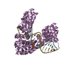
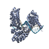
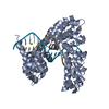
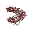
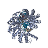
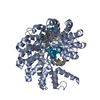
- PDB-4osi: Crystal structure of the TAL effector dHax3 with NI RVD at 2.8 an... -
+ Open data

- Basic information
| Entry | Database: PDB / ID: 4osi | ||||||
|---|---|---|---|---|---|---|---|
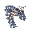
| Title | Crystal structure of the TAL effector dHax3 with NI RVD at 2.8 angstrom resolution | ||||||
 Components Components |
| ||||||
 Keywords Keywords | DNA binding protein/DNA / DNA binding protein / DNA / DNA binding protein-DNA complex | ||||||
| Function / homology | : / TAL effector repeat / TAL effector repeat / host cell nucleus / extracellular region / DNA / DNA (> 10) / Hax3 Function and homology information Function and homology information | ||||||
| Biological species |  Xanthomonas campestris pv. armoraciae (bacteria) Xanthomonas campestris pv. armoraciae (bacteria) | ||||||
| Method |  X-RAY DIFFRACTION / SYNCHROTRON / MOLECULAR REPLACEMENT / Resolution: 2.849 Å X-RAY DIFFRACTION / SYNCHROTRON / MOLECULAR REPLACEMENT / Resolution: 2.849 Å | ||||||
 Authors Authors | Deng, D. / Wu, J.P. / Yan, C.Y. / Pan, X.J. / Yan, N. | ||||||
 Citation Citation | Journal: Protein Cell / Year: 2014 Title: Revisiting the TALE repeat Authors: Deng, D. / Yan, C.Y. / Wu, J.P. / Pan, X.J. / Yan, N. | ||||||
| History |
|
- Structure visualization
| Structure viewer | Molecule: MolmilJmol/JSmol |
|---|
- Downloads & links
-Download
| PDBx/mmCIF format | 4osi.cif.gz | 429.3 KB | Display | PDBx/mmCIF format |
|---|---|---|---|---|
| PDB format | pdb4osi.ent.gz | 350.6 KB | Display | PDB format |
| PDBx/mmJSON format | 4osi.json.gz | Tree view | PDBx/mmJSON format | |
| Others |  Other downloads Other downloads |
-Validation report
| Arichive directory | https://data.pdbj.org/pub/pdb/validation_reports/os/4osiftp://data.pdbj.org/pub/pdb/validation_reports/os/4osi | HTTPS FTP |
|---|
-Related structure data
| Related structure data |  4oshC 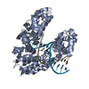 4osjC 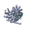 4oskC  4oslC 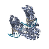 4osmC 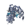 4osqC 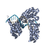 4osrC 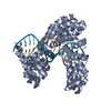 4ossC 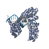 4ostC 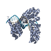 4osvC 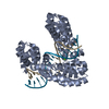 4oswC 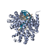 4oszC  4ot0C  4ot3C 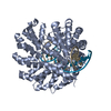 4otoC  3v6tS C: citing same article ( S: Starting model for refinement |
|---|---|
| Similar structure data |


-Links
 PDBj
PDBj


- Assembly
| Deposited unit | 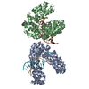
| ||||||||
|---|---|---|---|---|---|---|---|---|---|
| 1 | 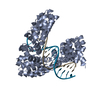
| ||||||||
| 2 | 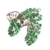
| ||||||||
| Unit cell |
|
-Components
| #1: Protein | Mass: 51780.586 Da / Num. of mol.: 2 / Fragment: UNP residues 231-720 Mutation: N300H,I301D,N368H,I369D,H402N,D403G,H436N,D437G,H470N,D471G,S505I,S539G,N572H,S573D,H606N,D607G,N640H,I641D Source method: isolated from a genetically manipulated source Source: (gene. exp.) Xanthomonas campestris pv. armoraciae (bacteria)Gene: hax3 / Production host: #2: DNA chain | Mass: 5070.281 Da / Num. of mol.: 2 / Source method: obtained synthetically #3: DNA chain | Mass: 5342.513 Da / Num. of mol.: 2 / Source method: obtained synthetically #4: Water | ChemComp-HOH / |  Mass: 18.015 Da / Num. of mol.: 39 / Source method: isolated from a natural source / Formula: H2O Mass: 18.015 Da / Num. of mol.: 39 / Source method: isolated from a natural source / Formula: H2O |
|---|
-Experimental details
-Experiment
| Experiment | Method: X-RAY DIFFRACTION / Number of used crystals: 1 |
|---|
- Sample preparation
| Crystal | Density Matthews: 2.66 Å3/Da / Density % sol: 53.67 % |
|---|---|
| Crystal grow | Temperature: 291 K / Method: vapor diffusion, hanging drop / pH: 6 Details: 10%-15% PEG 3350, 12% ethanol, 0.1M MES, pH 6.0, VAPOR DIFFUSION, HANGING DROP, temperature 291K |
-Data collection
| Diffraction | Mean temperature: 100 K |
|---|---|
| Diffraction source | Source: SYNCHROTRON / Site: SSRF  / Beamline: BL17U / Wavelength: 0.9792 Å / Beamline: BL17U / Wavelength: 0.9792 Å |
| Detector | Type: ADSC QUANTUM 315r / Detector: CCD / Date: Nov 8, 2011 |
| Radiation | Protocol: SINGLE WAVELENGTH / Monochromatic (M) / Laue (L): M / Scattering type: x-ray |
| Radiation wavelength | Wavelength: 0.9792 Å / Relative weight: 1 |
| Reflection | Resolution: 2.8→40 Å / Num. all: 32103 / Num. obs: 30448 / % possible obs: 94.8 % / Observed criterion σ(F): 2 / Observed criterion σ(I): 2 / Biso Wilson estimate: 75.59 Å2 |
- Processing
| Software |
| ||||||||||||||||||||||||||||||||||||||||||||||||||||||||||||||||||||||||||||||||||||
|---|---|---|---|---|---|---|---|---|---|---|---|---|---|---|---|---|---|---|---|---|---|---|---|---|---|---|---|---|---|---|---|---|---|---|---|---|---|---|---|---|---|---|---|---|---|---|---|---|---|---|---|---|---|---|---|---|---|---|---|---|---|---|---|---|---|---|---|---|---|---|---|---|---|---|---|---|---|---|---|---|---|---|---|---|---|
| Refinement | Method to determine structure: MOLECULAR REPLACEMENT Starting model: 3V6T Resolution: 2.849→31.986 Å / FOM work R set: 0.7055 / SU ML: 0.88 / σ(F): 1.34 / Phase error: 35.13 / Stereochemistry target values: ML
| ||||||||||||||||||||||||||||||||||||||||||||||||||||||||||||||||||||||||||||||||||||
| Solvent computation | Shrinkage radii: 1.06 Å / VDW probe radii: 1.3 Å / Solvent model: FLAT BULK SOLVENT MODEL / Bsol: 32.295 Å2 / ksol: 0.252 e/Å3 | ||||||||||||||||||||||||||||||||||||||||||||||||||||||||||||||||||||||||||||||||||||
| Displacement parameters | Biso max: 222.14 Å2 / Biso mean: 87.53 Å2 / Biso min: 42.34 Å2
| ||||||||||||||||||||||||||||||||||||||||||||||||||||||||||||||||||||||||||||||||||||
| Refinement step | Cycle: LAST / Resolution: 2.849→31.986 Å
| ||||||||||||||||||||||||||||||||||||||||||||||||||||||||||||||||||||||||||||||||||||
| Refine LS restraints |
| ||||||||||||||||||||||||||||||||||||||||||||||||||||||||||||||||||||||||||||||||||||
| LS refinement shell | Refine-ID: X-RAY DIFFRACTION / Total num. of bins used: 11
| ||||||||||||||||||||||||||||||||||||||||||||||||||||||||||||||||||||||||||||||||||||
| Refinement TLS params. | Method: refined / Origin x: -14.8927 Å / Origin y: -36.1106 Å / Origin z: -25.8037 Å
| ||||||||||||||||||||||||||||||||||||||||||||||||||||||||||||||||||||||||||||||||||||
| Refinement TLS group | Selection details: ALL |
