 Movie
Movie Controller


+ Open data

- Basic information
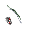
| Entry | Database: PDB / ID: 4onk | ||||||
|---|---|---|---|---|---|---|---|
| Title | [Leu-5]-Enkephalin mutant - YVVFL | ||||||
 Components Components | [Leu-5]-Enkephalin mutant - YVVFL | ||||||
 Keywords Keywords | PROTEIN FIBRIL / amyloid-like protofibril | ||||||
| Biological species | synthetic construct (others) | ||||||
| Method |  X-RAY DIFFRACTION / SYNCHROTRON / MOLECULAR REPLACEMENT / molecular replacement / Resolution: 1.9 Å X-RAY DIFFRACTION / SYNCHROTRON / MOLECULAR REPLACEMENT / molecular replacement / Resolution: 1.9 Å | ||||||
 Authors Authors | Sangwan, S. / Eisenberg, D. / Sawaya, M.R. / Do, T.D. / Bowers, M.T. / Lapointe, N.E. / Teplow, D.B. / Feinstein, S.C. | ||||||
 Citation Citation | Journal: J.Phys.Chem.B / Year: 2014 Title: Factors that drive Peptide assembly from native to amyloid structures: experimental and theoretical analysis of [leu-5]-enkephalin mutants. Authors: Do, T.D. / LaPointe, N.E. / Sangwan, S. / Teplow, D.B. / Feinstein, S.C. / Sawaya, M.R. / Eisenberg, D.S. / Bowers, M.T. | ||||||
| History |
|
- Structure visualization
| Structure viewer | Molecule:  MolmilJmol/JSmol MolmilJmol/JSmol |
|---|
- Downloads & links
-Download
| PDBx/mmCIF format | 4onk.cif.gz | 9.9 KB | Display | PDBx/mmCIF format |
|---|---|---|---|---|
| PDB format | pdb4onk.ent.gz | 5.5 KB | Display | PDB format |
| PDBx/mmJSON format | 4onk.json.gz | Tree view | PDBx/mmJSON format | |
| Others |  Other downloads Other downloads |
-Validation report
| Arichive directory | https://data.pdbj.org/pub/pdb/validation_reports/on/4onkftp://data.pdbj.org/pub/pdb/validation_reports/on/4onk | HTTPS FTP |
|---|
-Related structure data

-Links
 PDBj
PDBj- Assembly
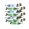
| Deposited unit | 
| ||||||||
|---|---|---|---|---|---|---|---|---|---|
| 1 | x 6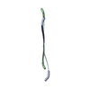
| ||||||||
| Unit cell |
| ||||||||
| Details | The biological unit is a pair of beta sheets. One sheet is constructed from chains A and B with unit cell translations along the a direction (i.e. X+1,Y,Z; X+2,Y,Z; X+3,Y,Z, etc.). The second sheet is constructed from X,Y+1,Z; X+1,Y+1,Z; X+2,Y+1,Z; etc.). |
-Components
| #1: Protein/peptide | Mass: 639.782 Da / Num. of mol.: 2 / Source method: obtained synthetically / Details: synthesized / Source: (synth.) synthetic construct (others) #2: Water | ChemComp-HOH / |  Mass: 18.015 Da / Num. of mol.: 3 / Source method: isolated from a natural source / Formula: H2O Mass: 18.015 Da / Num. of mol.: 3 / Source method: isolated from a natural source / Formula: H2O |
|---|
-Experimental details
-Experiment
| Experiment | Method: X-RAY DIFFRACTION / Number of used crystals: 3 |
|---|
- Sample preparation
| Crystal grow | Temperature: 298 K / Method: vapor diffusion, hanging drop Details: reservoir contained 25% PEG 3350, 0.2M Potassium Thiocyanate, vapor diffusion, hanging drop, temperature 298K |
|---|
-Data collection
| Diffraction | Mean temperature: 100 K | |||||||||||||||||||||||||||||||||||||||||||||||||||||||||||||||||||||||||||||
|---|---|---|---|---|---|---|---|---|---|---|---|---|---|---|---|---|---|---|---|---|---|---|---|---|---|---|---|---|---|---|---|---|---|---|---|---|---|---|---|---|---|---|---|---|---|---|---|---|---|---|---|---|---|---|---|---|---|---|---|---|---|---|---|---|---|---|---|---|---|---|---|---|---|---|---|---|---|---|
| Diffraction source | Source: SYNCHROTRON / Site: APS  / Beamline: 24-ID-E / Wavelength: 0.9791 Å / Beamline: 24-ID-E / Wavelength: 0.9791 Å | |||||||||||||||||||||||||||||||||||||||||||||||||||||||||||||||||||||||||||||
| Detector | Type: ADSC QUANTUM 315 / Detector: CCD / Date: Aug 8, 2013 | |||||||||||||||||||||||||||||||||||||||||||||||||||||||||||||||||||||||||||||
| Radiation | Protocol: SINGLE WAVELENGTH / Monochromatic (M) / Laue (L): M / Scattering type: x-ray | |||||||||||||||||||||||||||||||||||||||||||||||||||||||||||||||||||||||||||||
| Radiation wavelength | Wavelength: 0.9791 Å / Relative weight: 1 | |||||||||||||||||||||||||||||||||||||||||||||||||||||||||||||||||||||||||||||
| Reflection | Resolution: 1.9→100 Å / Num. all: 537 / Num. obs: 537 / % possible obs: 96.9 % / Observed criterion σ(I): -3 / Redundancy: 3.8 % / Biso Wilson estimate: 8.11 Å2 / Rmerge(I) obs: 0.151 / Χ2: 3.634 / Net I/σ(I): 10.7 | |||||||||||||||||||||||||||||||||||||||||||||||||||||||||||||||||||||||||||||
| Reflection shell |
|
-Phasing
| Phasing | Method: molecular replacement | |||||||||
|---|---|---|---|---|---|---|---|---|---|---|
| Phasing MR | Model details: Phaser MODE: MR_AUTO
|
- Processing
| Software |
| |||||||||||||||||||||||||
|---|---|---|---|---|---|---|---|---|---|---|---|---|---|---|---|---|---|---|---|---|---|---|---|---|---|---|
| Refinement | Method to determine structure: MOLECULAR REPLACEMENT Starting model: ideal beta strand with sequence AVVAA Resolution: 1.9→9.278 Å / FOM work R set: 0.8404 / SU ML: 0.29 / σ(F): 35.13 / Phase error: 23.66 / Stereochemistry target values: LS_WUNIT_K1
| |||||||||||||||||||||||||
| Solvent computation | Shrinkage radii: 0.9 Å / VDW probe radii: 1.11 Å / Solvent model: FLAT BULK SOLVENT MODEL | |||||||||||||||||||||||||
| Displacement parameters | Biso max: 22.76 Å2 / Biso mean: 14.15 Å2 / Biso min: 8.29 Å2 | |||||||||||||||||||||||||
| Refinement step | Cycle: LAST / Resolution: 1.9→9.278 Å /
| |||||||||||||||||||||||||
| Refine LS restraints |
| |||||||||||||||||||||||||
| LS refinement shell | Highest resolution: 1.9003 Å / Total num. of bins used: 1
|
