 Movie
Movie Controller

[English] 日本語
 Yorodumi
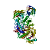
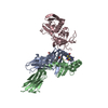
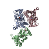
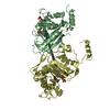
- PDB-4okv: Crystal structure of anopheline anti-platelet protein with Fab an... -
+ Open data

- Basic information
| Entry | Database: PDB / ID: 4okv | ||||||
|---|---|---|---|---|---|---|---|
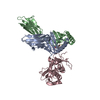
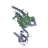
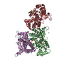
| Title | Crystal structure of anopheline anti-platelet protein with Fab antibody | ||||||
 Components Components |
| ||||||
 Keywords Keywords | BLOOD CLOTTING / malaria vector mosquito / collagen binding protein | ||||||
| Function / homology |  Function and homology information Function and homology information | ||||||
| Biological species |   | ||||||
| Method |  X-RAY DIFFRACTION / SYNCHROTRON / MOLECULAR REPLACEMENT / molecular replacement / Resolution: 1.802 Å X-RAY DIFFRACTION / SYNCHROTRON / MOLECULAR REPLACEMENT / molecular replacement / Resolution: 1.802 Å | ||||||
 Authors Authors | Park, S.Y. / Sugiyama, K. | ||||||
 Citation Citation | Journal: To be Published Title: The crystal structure of anopheles anti-platelet protein, a powerful anti-coagulant, in complex with an antibody Authors: Sugiyama, K. / Iyori, M. / Sawaguchi, A. / Akashi, S. / Tame, J.R.H. / Yoshida, S. / Park, S.Y. | ||||||
| History |
|
- Structure visualization
| Structure viewer | Molecule: MolmilJmol/JSmol |
|---|
- Downloads & links
-Download
| PDBx/mmCIF format | 4okv.cif.gz | 213.7 KB | Display | PDBx/mmCIF format |
|---|---|---|---|---|
| PDB format | pdb4okv.ent.gz | 170.4 KB | Display | PDB format |
| PDBx/mmJSON format | 4okv.json.gz | Tree view | PDBx/mmJSON format | |
| Others |  Other downloads Other downloads |
-Validation report
| Summary document | 4okv_validation.pdf.gz | 458.1 KB | Display | wwPDB validaton report |
|---|---|---|---|---|
| Full document | 4okv_full_validation.pdf.gz | 462.9 KB | Display | |
| Data in XML | 4okv_validation.xml.gz | 41.6 KB | Display | |
| Data in CIF | 4okv_validation.cif.gz | 61.3 KB | Display | |
| Arichive directory | https://data.pdbj.org/pub/pdb/validation_reports/ok/4okvftp://data.pdbj.org/pub/pdb/validation_reports/ok/4okv | HTTPS FTP |
-Related structure data
| Similar structure data |
|---|

-Links
 PDBj
PDBj

- Assembly
| Deposited unit | 
| ||||||||
|---|---|---|---|---|---|---|---|---|---|
| 1 | 
| ||||||||
| 2 | 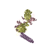
| ||||||||
| Unit cell |
|
-Components
| #1: Antibody | Mass: 23085.854 Da / Num. of mol.: 2 / Source method: isolated from a natural source / Details: hybridoma cells / Source: (natural) #2: Antibody | Mass: 24015.744 Da / Num. of mol.: 2 / Source method: isolated from a natural source / Details: hybridoma cells / Source: (natural) #3: Protein | Mass: 7584.399 Da / Num. of mol.: 2 / Fragment: UNP residues 201-266 Source method: isolated from a genetically manipulated source Source: (gene. exp.) Gene: ansg-1, aapp / Plasmid: pET22 / Production host:  #4: Water | ChemComp-HOH / |  Mass: 18.015 Da / Num. of mol.: 698 / Source method: isolated from a natural source / Formula: H2O Mass: 18.015 Da / Num. of mol.: 698 / Source method: isolated from a natural source / Formula: H2OHas protein modification | Y | |
|---|
-Experimental details
-Experiment
| Experiment | Method: X-RAY DIFFRACTION / Number of used crystals: 1 |
|---|
- Sample preparation
| Crystal | Density Matthews: 3.54 Å3/Da / Density % sol: 65.25 % / Mosaicity: 0.281 ° |
|---|---|
| Crystal grow | Temperature: 298 K / Method: vapor diffusion, hanging drop / pH: 7 Details: 0.1M HEPES, 15% PEG 20000, pH 7.0, vapor diffusion, hanging drop, temperature 298K |
-Data collection
| Diffraction | Mean temperature: 298 K | ||||||||||||||||||||||||||||||||||||||||||||||||||||||||||||||||||||||||||||||||||||||||||||||||||||||||||||||||||||||||||||||
|---|---|---|---|---|---|---|---|---|---|---|---|---|---|---|---|---|---|---|---|---|---|---|---|---|---|---|---|---|---|---|---|---|---|---|---|---|---|---|---|---|---|---|---|---|---|---|---|---|---|---|---|---|---|---|---|---|---|---|---|---|---|---|---|---|---|---|---|---|---|---|---|---|---|---|---|---|---|---|---|---|---|---|---|---|---|---|---|---|---|---|---|---|---|---|---|---|---|---|---|---|---|---|---|---|---|---|---|---|---|---|---|---|---|---|---|---|---|---|---|---|---|---|---|---|---|---|---|
| Diffraction source | Source: SYNCHROTRON / Site: Photon Factory  / Beamline: BL-17A / Wavelength: 0.98 Å / Beamline: BL-17A / Wavelength: 0.98 Å | ||||||||||||||||||||||||||||||||||||||||||||||||||||||||||||||||||||||||||||||||||||||||||||||||||||||||||||||||||||||||||||||
| Detector | Type: ADSC QUANTUM 315 / Detector: CCD / Date: Jul 6, 2011 | ||||||||||||||||||||||||||||||||||||||||||||||||||||||||||||||||||||||||||||||||||||||||||||||||||||||||||||||||||||||||||||||
| Radiation | Monochromator: Si 111 CHANNEL / Protocol: SINGLE WAVELENGTH / Monochromatic (M) / Laue (L): M / Scattering type: x-ray | ||||||||||||||||||||||||||||||||||||||||||||||||||||||||||||||||||||||||||||||||||||||||||||||||||||||||||||||||||||||||||||||
| Radiation wavelength | Wavelength: 0.98 Å / Relative weight: 1 | ||||||||||||||||||||||||||||||||||||||||||||||||||||||||||||||||||||||||||||||||||||||||||||||||||||||||||||||||||||||||||||||
| Reflection | Resolution: 1.8→50 Å / Num. obs: 135319 / % possible obs: 94.5 % / Redundancy: 3.9 % / Biso Wilson estimate: 17.15 Å2 / Rmerge(I) obs: 0.093 / Χ2: 1.499 / Net I/σ(I): 9.6 | ||||||||||||||||||||||||||||||||||||||||||||||||||||||||||||||||||||||||||||||||||||||||||||||||||||||||||||||||||||||||||||||
| Reflection shell | Diffraction-ID: 1 / Rejects: _
|
-Phasing
| Phasing | Method: molecular replacement |
|---|
- Processing
| Software |
| |||||||||||||||||||||||||||||||||||||||||||||||||||||||||||||||||||||||||||||||||||||||||||||||||||||||||||||||||||||||||||||||||||||||||||||||||||||||||||||||||||||||||||||||||||||||||||||||||||||||||||||||||||||||||
|---|---|---|---|---|---|---|---|---|---|---|---|---|---|---|---|---|---|---|---|---|---|---|---|---|---|---|---|---|---|---|---|---|---|---|---|---|---|---|---|---|---|---|---|---|---|---|---|---|---|---|---|---|---|---|---|---|---|---|---|---|---|---|---|---|---|---|---|---|---|---|---|---|---|---|---|---|---|---|---|---|---|---|---|---|---|---|---|---|---|---|---|---|---|---|---|---|---|---|---|---|---|---|---|---|---|---|---|---|---|---|---|---|---|---|---|---|---|---|---|---|---|---|---|---|---|---|---|---|---|---|---|---|---|---|---|---|---|---|---|---|---|---|---|---|---|---|---|---|---|---|---|---|---|---|---|---|---|---|---|---|---|---|---|---|---|---|---|---|---|---|---|---|---|---|---|---|---|---|---|---|---|---|---|---|---|---|---|---|---|---|---|---|---|---|---|---|---|---|---|---|---|---|---|---|---|---|---|---|---|---|---|---|---|---|---|---|---|---|
| Refinement | Method to determine structure: MOLECULAR REPLACEMENT / Resolution: 1.802→47.624 Å / FOM work R set: 0.8172 / SU ML: 0.25 / σ(F): 1.42 / Phase error: 25.73 / Stereochemistry target values: ML
| |||||||||||||||||||||||||||||||||||||||||||||||||||||||||||||||||||||||||||||||||||||||||||||||||||||||||||||||||||||||||||||||||||||||||||||||||||||||||||||||||||||||||||||||||||||||||||||||||||||||||||||||||||||||||
| Solvent computation | Shrinkage radii: 1.11 Å / VDW probe radii: 1.3 Å / Solvent model: FLAT BULK SOLVENT MODEL / Bsol: 21.356 Å2 / ksol: 0.322 e/Å3 | |||||||||||||||||||||||||||||||||||||||||||||||||||||||||||||||||||||||||||||||||||||||||||||||||||||||||||||||||||||||||||||||||||||||||||||||||||||||||||||||||||||||||||||||||||||||||||||||||||||||||||||||||||||||||
| Displacement parameters | Biso max: 104.5 Å2 / Biso mean: 25.16 Å2 / Biso min: 9.12 Å2
| |||||||||||||||||||||||||||||||||||||||||||||||||||||||||||||||||||||||||||||||||||||||||||||||||||||||||||||||||||||||||||||||||||||||||||||||||||||||||||||||||||||||||||||||||||||||||||||||||||||||||||||||||||||||||
| Refinement step | Cycle: LAST / Resolution: 1.802→47.624 Å
| |||||||||||||||||||||||||||||||||||||||||||||||||||||||||||||||||||||||||||||||||||||||||||||||||||||||||||||||||||||||||||||||||||||||||||||||||||||||||||||||||||||||||||||||||||||||||||||||||||||||||||||||||||||||||
| Refine LS restraints |
| |||||||||||||||||||||||||||||||||||||||||||||||||||||||||||||||||||||||||||||||||||||||||||||||||||||||||||||||||||||||||||||||||||||||||||||||||||||||||||||||||||||||||||||||||||||||||||||||||||||||||||||||||||||||||
| LS refinement shell | Refine-ID: X-RAY DIFFRACTION / Total num. of bins used: 30
|
