 ムービー
ムービー コントローラー


+ データを開く

- 基本情報
基本情報
| 登録情報 | データベース: PDB / ID: 4o5a | ||||||
|---|---|---|---|---|---|---|---|
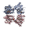
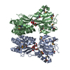
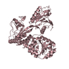
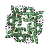
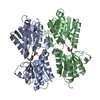
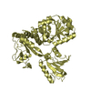
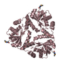
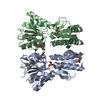
| タイトル | The crystal structure of a LacI family transcriptional regulator from Bifidobacterium animalis subsp. lactis DSM 10140 | ||||||
 要素 要素 | LacI family transcription regulator | ||||||
 キーワード キーワード | TRANSCRIPTION REGULATOR / structural genomics / PSI-Biology / protein structure initiative / midwest center for structural genomics / MCSG | ||||||
| 機能・相同性 | Response regulator / Rossmann fold / 3-Layer(aba) Sandwich / Alpha Beta / :  機能・相同性情報 機能・相同性情報 | ||||||
| 生物種 |  Bifidobacterium animalis subsp. lactis (バクテリア) Bifidobacterium animalis subsp. lactis (バクテリア) | ||||||
| 手法 |  X線回折 / シンクロトロン / 単波長異常分散 / 解像度: 1.777 Å X線回折 / シンクロトロン / 単波長異常分散 / 解像度: 1.777 Å | ||||||
 データ登録者 データ登録者 | Tan, K. / Li, H. / Endres, M. / Joachimiak, A. / Midwest Center for Structural Genomics (MCSG) | ||||||
 引用 引用 | ジャーナル: To be Published タイトル: The crystal structure of a LacI family transcriptional regulator from Bifidobacterium animalis subsp. lactis DSM 10140. 著者: Tan, K. / Li, H. / Endres, M. / Joachimiak, A. | ||||||
| 履歴 |
|
- 構造の表示
構造の表示
| 構造ビューア | 分子: MolmilJmol/JSmol |
|---|
- ダウンロードとリンク
-ダウンロード
| PDBx/mmCIF形式 | 4o5a.cif.gz | 125 KB | 表示 | PDBx/mmCIF形式 |
|---|---|---|---|---|
| PDB形式 | pdb4o5a.ent.gz | 97.3 KB | 表示 | PDB形式 |
| PDBx/mmJSON形式 | 4o5a.json.gz | ツリー表示 | PDBx/mmJSON形式 | |
| その他 |  その他のダウンロード その他のダウンロード |
-検証レポート
| アーカイブディレクトリ | https://data.pdbj.org/pub/pdb/validation_reports/o5/4o5aftp://data.pdbj.org/pub/pdb/validation_reports/o5/4o5a | HTTPS FTP |
|---|
-関連構造データ
| 類似構造データ | |
|---|---|
| その他のデータベース |
-リンク
 PDBj
PDBj- 集合体
集合体
| 登録構造単位 | 
| |||||||||
|---|---|---|---|---|---|---|---|---|---|---|
| 1 | 
| |||||||||
| 単位格子 |
| |||||||||
| Components on special symmetry positions |
| |||||||||
| 詳細 | Experimentally unknown. It is predicted that the chain A and its symmetry-related molecule by an operator (-x+1,y,-z+1) form a dimer. |
-要素
| #1: タンパク質 | 分子量: 37823.988 Da / 分子数: 1 / 由来タイプ: 組換発現 由来: (組換発現) Bifidobacterium animalis subsp. lactis (バクテリア)株: DSM 10140 / 遺伝子: Balat_0153 / プラスミド: pMCSG68 / 発現宿主: | ||||||
|---|---|---|---|---|---|---|---|
| #2: 化合物 |   分子量: 92.094 Da / 分子数: 3 / 由来タイプ: 合成 / 式: C3H8O3 分子量: 92.094 Da / 分子数: 3 / 由来タイプ: 合成 / 式: C3H8O3#3: 化合物 |   分子量: 96.063 Da / 分子数: 2 / 由来タイプ: 合成 / 式: SO4 分子量: 96.063 Da / 分子数: 2 / 由来タイプ: 合成 / 式: SO4#4: 水 | ChemComp-HOH / |  分子量: 18.015 Da / 分子数: 195 / 由来タイプ: 天然 / 式: H2O 分子量: 18.015 Da / 分子数: 195 / 由来タイプ: 天然 / 式: H2OHas protein modification | Y | |
-実験情報
-実験
| 実験 | 手法: X線回折 / 使用した結晶の数: 1 |
|---|
- 試料調製
試料調製
| 結晶 | マシュー密度: 2.31 Å3/Da / 溶媒含有率: 46.8 % |
|---|---|
| 結晶化 | 温度: 289 K / 手法: 蒸気拡散法, シッティングドロップ法 / pH: 8.5 詳細: 0.2M Ammonium Sulfate, 0.1M Tris:HCl, 25% (w/v) PEG3350, pH 8.5, VAPOR DIFFUSION, SITTING DROP, temperature 289K |
-データ収集
| 回折 | 平均測定温度: 100 K |
|---|---|
| 放射光源 | 由来: シンクロトロン / サイト: APS  / ビームライン: 19-ID / 波長: 0.97921 Å / ビームライン: 19-ID / 波長: 0.97921 Å |
| 検出器 | タイプ: ADSC QUANTUM 210r / 検出器: CCD / 日付: 2013年11月27日 / 詳細: mirror |
| 放射 | モノクロメーター: Si 111 crystal / プロトコル: SINGLE WAVELENGTH / 単色(M)・ラウエ(L): M / 散乱光タイプ: x-ray |
| 放射波長 | 波長: 0.97921 Å / 相対比: 1 |
| 反射 | 解像度: 1.78→27 Å / Num. all: 32091 / Num. obs: 32091 / % possible obs: 97.1 % / Observed criterion σ(F): 0 / Observed criterion σ(I): -5 / 冗長度: 4.7 % / Biso Wilson estimate: 23.31 Å2 / Rmerge(I) obs: 0.101 / Net I/σ(I): 33.4 |
| 反射 シェル | 解像度: 1.78→1.81 Å / 冗長度: 4.8 % / Rmerge(I) obs: 0.751 / Mean I/σ(I) obs: 2.5 / Num. unique all: 1576 / % possible all: 96.9 |
- 解析
解析
| ソフトウェア |
| ||||||||||||||||||||||||||||||||||||||||||||||||||||||||||||||||||||||||||||||||||||||||||||||||||||
|---|---|---|---|---|---|---|---|---|---|---|---|---|---|---|---|---|---|---|---|---|---|---|---|---|---|---|---|---|---|---|---|---|---|---|---|---|---|---|---|---|---|---|---|---|---|---|---|---|---|---|---|---|---|---|---|---|---|---|---|---|---|---|---|---|---|---|---|---|---|---|---|---|---|---|---|---|---|---|---|---|---|---|---|---|---|---|---|---|---|---|---|---|---|---|---|---|---|---|---|---|---|
| 精密化 | 構造決定の手法: 単波長異常分散 / 解像度: 1.777→26.967 Å / SU ML: 0.18 / σ(F): 1.36 / 位相誤差: 21.38 / 立体化学のターゲット値: ML
| ||||||||||||||||||||||||||||||||||||||||||||||||||||||||||||||||||||||||||||||||||||||||||||||||||||
| 溶媒の処理 | 減衰半径: 0.9 Å / VDWプローブ半径: 1.11 Å / 溶媒モデル: FLAT BULK SOLVENT MODEL | ||||||||||||||||||||||||||||||||||||||||||||||||||||||||||||||||||||||||||||||||||||||||||||||||||||
| 精密化ステップ | サイクル: LAST / 解像度: 1.777→26.967 Å
| ||||||||||||||||||||||||||||||||||||||||||||||||||||||||||||||||||||||||||||||||||||||||||||||||||||
| 拘束条件 |
| ||||||||||||||||||||||||||||||||||||||||||||||||||||||||||||||||||||||||||||||||||||||||||||||||||||
| LS精密化 シェル |
| ||||||||||||||||||||||||||||||||||||||||||||||||||||||||||||||||||||||||||||||||||||||||||||||||||||
| 精密化 TLS | 手法: refined / Refine-ID: X-RAY DIFFRACTION
| ||||||||||||||||||||||||||||||||||||||||||||||||||||||||||||||||||||||||||||||||||||||||||||||||||||
| 精密化 TLSグループ |
|
