 Movie
Movie Controller

[English] 日本語
 Yorodumi
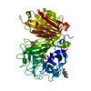
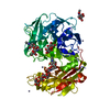
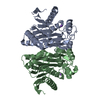
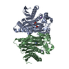
- PDB-4o0e: Crystal structure of the human L-asparaginase protein T186V mutant -
+ Open data

- Basic information
| Entry | Database: PDB / ID: 4o0e | ||||||
|---|---|---|---|---|---|---|---|
| Title | Crystal structure of the human L-asparaginase protein T186V mutant | ||||||
 Components Components | Isoaspartyl peptidase/L-asparaginase | ||||||
 Keywords Keywords | HYDROLASE / NTN ENZYME / HOMODIMER / asparaginase | ||||||
| Function / homology |  Function and homology information Function and homology informationasparagine catabolic process via L-aspartate / beta-aspartyl-peptidase / Phenylalanine metabolism / asparaginase / asparaginase activity / beta-aspartyl-peptidase activity / photoreceptor inner segment / proteolysis / nucleus / cytosol / cytoplasm Similarity search - Function | ||||||
| Biological species |  Homo sapiens (human) Homo sapiens (human) | ||||||
| Method |  X-RAY DIFFRACTION / SYNCHROTRON / MOLECULAR REPLACEMENT / Resolution: 1.71 Å X-RAY DIFFRACTION / SYNCHROTRON / MOLECULAR REPLACEMENT / Resolution: 1.71 Å | ||||||
 Authors Authors | Nomme, J. / Lavie, A. | ||||||
 Citation Citation | Journal: J.Mol.Biol. / Year: 2014 Title: Elucidation of the Specific Function of the Conserved Threonine Triad Responsible for Human l-Asparaginase Autocleavage and Substrate Hydrolysis. Authors: Nomme, J. / Su, Y. / Lavie, A. | ||||||
| History |
|
- Structure visualization
| Structure viewer | Molecule: MolmilJmol/JSmol |
|---|
- Downloads & links
-Download
| PDBx/mmCIF format | 4o0e.cif.gz | 123 KB | Display | PDBx/mmCIF format |
|---|---|---|---|---|
| PDB format | pdb4o0e.ent.gz | 94.8 KB | Display | PDB format |
| PDBx/mmJSON format | 4o0e.json.gz | Tree view | PDBx/mmJSON format | |
| Others |  Other downloads Other downloads |
-Validation report
| Summary document | 4o0e_validation.pdf.gz | 437.6 KB | Display | wwPDB validaton report |
|---|---|---|---|---|
| Full document | 4o0e_full_validation.pdf.gz | 445.6 KB | Display | |
| Data in XML | 4o0e_validation.xml.gz | 24 KB | Display | |
| Data in CIF | 4o0e_validation.cif.gz | 33.8 KB | Display | |
| Arichive directory | https://data.pdbj.org/pub/pdb/validation_reports/o0/4o0eftp://data.pdbj.org/pub/pdb/validation_reports/o0/4o0e | HTTPS FTP |
-Related structure data
| Related structure data |  4o0cC  4o0dC  4o0fC  4o0gC  4o0hC  4gdv C: citing same article ( S: Starting model for refinement |
|---|---|
| Similar structure data |

-Links
 PDBj
PDBj


- Assembly
| Deposited unit | 
| ||||||||
|---|---|---|---|---|---|---|---|---|---|
| 1 |
| ||||||||
| Unit cell |
|
-Components
| #1: Protein | Mass: 32230.666 Da / Num. of mol.: 2 / Mutation: T186V Source method: isolated from a genetically manipulated source Source: (gene. exp.) Homo sapiens (human) / Gene: ASRGL1, ALP, CRASH / Plasmid: pET14b / Production host:  References: UniProt: Q7L266, beta-aspartyl-peptidase, asparaginase #2: Chemical |   Mass: 22.990 Da / Num. of mol.: 2 / Source method: obtained synthetically / Formula: Na Mass: 22.990 Da / Num. of mol.: 2 / Source method: obtained synthetically / Formula: Na#3: Water | ChemComp-HOH / |  Mass: 18.015 Da / Num. of mol.: 177 / Source method: isolated from a natural source / Formula: H2O Mass: 18.015 Da / Num. of mol.: 177 / Source method: isolated from a natural source / Formula: H2O |
|---|
-Experimental details
-Experiment
| Experiment | Method: X-RAY DIFFRACTION / Number of used crystals: 1 |
|---|
- Sample preparation
| Crystal | Density Matthews: 2.36 Å3/Da / Density % sol: 47.77 % |
|---|---|
| Crystal grow | Temperature: 293 K / Method: vapor diffusion, hanging drop / pH: 7 Details: 2.2-2.5 M sodium Malonate, pH 7.0, VAPOR DIFFUSION, HANGING DROP, temperature 293K |
-Data collection
| Diffraction | Mean temperature: 100 K | |||||||||||||||
|---|---|---|---|---|---|---|---|---|---|---|---|---|---|---|---|---|
| Diffraction source | Source: SYNCHROTRON / Site: APS  / Beamline: 21-ID-D / Wavelength: 1.0782 Å / Beamline: 21-ID-D / Wavelength: 1.0782 Å | |||||||||||||||
| Detector | Type: MARMOSAIC 300 mm CCD / Detector: CCD / Date: Oct 11, 2012 | |||||||||||||||
| Radiation | Monochromator: C(111) / Protocol: SINGLE WAVELENGTH / Monochromatic (M) / Laue (L): M / Scattering type: x-ray | |||||||||||||||
| Radiation wavelength | Wavelength: 1.0782 Å / Relative weight: 1 | |||||||||||||||
| Reflection twin |
| |||||||||||||||
| Reflection | Resolution: 1.71→30 Å / Num. all: 62537 / Num. obs: 62537 / % possible obs: 98.1 % / Observed criterion σ(F): 0 / Observed criterion σ(I): 0 / Redundancy: 5.28 % / Rsym value: 0.062 / Net I/σ(I): 14.41 | |||||||||||||||
| Reflection shell | Resolution: 1.71→1.81 Å / Redundancy: 3.3 % / Mean I/σ(I) obs: 1.65 / Rsym value: 0.559 / % possible all: 93.1 |
- Processing
| Software |
| |||||||||||||||||||||||||||||||||||||||||||||||||||||||||||||||||||||||||||||||||||||||||||||||||||||||||
|---|---|---|---|---|---|---|---|---|---|---|---|---|---|---|---|---|---|---|---|---|---|---|---|---|---|---|---|---|---|---|---|---|---|---|---|---|---|---|---|---|---|---|---|---|---|---|---|---|---|---|---|---|---|---|---|---|---|---|---|---|---|---|---|---|---|---|---|---|---|---|---|---|---|---|---|---|---|---|---|---|---|---|---|---|---|---|---|---|---|---|---|---|---|---|---|---|---|---|---|---|---|---|---|---|---|---|
| Refinement | Method to determine structure: MOLECULAR REPLACEMENT Starting model: PDB entry 4GDV 4gdv Resolution: 1.71→28.45 Å / Cor.coef. Fo:Fc: 0.964 / Cor.coef. Fo:Fc free: 0.94 / SU B: 1.113 / SU ML: 0.041 / Cross valid method: THROUGHOUT / σ(F): 0 / ESU R: 0.022 / ESU R Free: 0.024 / Stereochemistry target values: MAXIMUM LIKELIHOOD / Details: HYDROGENS HAVE BEEN ADDED IN THE RIDING POSITIONS
| |||||||||||||||||||||||||||||||||||||||||||||||||||||||||||||||||||||||||||||||||||||||||||||||||||||||||
| Solvent computation | Ion probe radii: 0.8 Å / Shrinkage radii: 0.8 Å / VDW probe radii: 1.2 Å / Solvent model: MASK | |||||||||||||||||||||||||||||||||||||||||||||||||||||||||||||||||||||||||||||||||||||||||||||||||||||||||
| Displacement parameters | Biso mean: 26.426 Å2
| |||||||||||||||||||||||||||||||||||||||||||||||||||||||||||||||||||||||||||||||||||||||||||||||||||||||||
| Refinement step | Cycle: LAST / Resolution: 1.71→28.45 Å
| |||||||||||||||||||||||||||||||||||||||||||||||||||||||||||||||||||||||||||||||||||||||||||||||||||||||||
| Refine LS restraints |
| |||||||||||||||||||||||||||||||||||||||||||||||||||||||||||||||||||||||||||||||||||||||||||||||||||||||||
| LS refinement shell | Resolution: 1.71→1.755 Å / Total num. of bins used: 20
|
