 Movie
Movie Controller

[English] 日本語
 Yorodumi
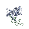
- PDB-4ngc: Structure of human Dicer Platform-PAZ-Connector Helix cassette in... -
+ Open data

- Basic information
| Entry | Database: PDB / ID: 4ngc | ||||||
|---|---|---|---|---|---|---|---|
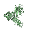
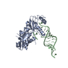
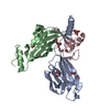
| Title | Structure of human Dicer Platform-PAZ-Connector Helix cassette in complex with 12-mer siRNA having UA-3' ends (2.1 Angstrom resolution) | ||||||
 Components Components |
| ||||||
 Keywords Keywords | HYDROLASE/RNA / PAZ domain / platform domain / connector helix / siRNA / RNase III domain / endoribonuclease / pre-miRNA / HYDROLASE-RNA complex | ||||||
| Function / homology |  Function and homology information Function and homology informationperipheral nervous system myelin formation / positive regulation of Schwann cell differentiation / global gene silencing by mRNA cleavage / negative regulation of Schwann cell proliferation / tRNA-derived small RNA (tsRNA or tRNA-related fragment, tRF) biogenesis / pre-miRNA binding / tRNA decay / ribonuclease III / Small interfering RNA (siRNA) biogenesis / apoptotic DNA fragmentation ...peripheral nervous system myelin formation / positive regulation of Schwann cell differentiation / global gene silencing by mRNA cleavage / negative regulation of Schwann cell proliferation / tRNA-derived small RNA (tsRNA or tRNA-related fragment, tRF) biogenesis / pre-miRNA binding / tRNA decay / ribonuclease III / Small interfering RNA (siRNA) biogenesis / apoptotic DNA fragmentation / deoxyribonuclease I activity / nerve development / positive regulation of myelination / RISC-loading complex / miRNA metabolic process / ribonuclease III activity / RISC complex assembly / miRNA processing / pre-miRNA processing / siRNA binding / M-decay: degradation of maternal mRNAs by maternally stored factors / siRNA processing / Regulation of MITF-M-dependent genes involved in apoptosis / RISC complex / MicroRNA (miRNA) biogenesis / negative regulation of tumor necrosis factor production / negative regulation of tumor necrosis factor-mediated signaling pathway / neuron projection morphogenesis / RNA endonuclease activity / helicase activity / double-stranded RNA binding / protein domain specific binding / negative regulation of gene expression / perinuclear region of cytoplasm / negative regulation of transcription by RNA polymerase II / RNA binding / extracellular exosome / ATP binding / metal ion binding / nucleus / cytoplasm / cytosol Similarity search - Function | ||||||
| Biological species |  Homo sapiens (human) Homo sapiens (human) | ||||||
| Method |  X-RAY DIFFRACTION / SYNCHROTRON / MOLECULAR REPLACEMENT / molecular replacement / Resolution: 2.104 Å X-RAY DIFFRACTION / SYNCHROTRON / MOLECULAR REPLACEMENT / molecular replacement / Resolution: 2.104 Å | ||||||
 Authors Authors | Simanshu, D.K. / Tian, Y. / Patel, D.J. | ||||||
 Citation Citation | Journal: Mol.Cell / Year: 2014 Title: A Phosphate-Binding Pocket within the Platform-PAZ-Connector Helix Cassette of Human Dicer. Authors: Tian, Y. / Simanshu, D.K. / Ma, J.B. / Park, J.E. / Heo, I. / Kim, V.N. / Patel, D.J. | ||||||
| History |
|
- Structure visualization
| Structure viewer | Molecule: MolmilJmol/JSmol |
|---|
- Downloads & links
-Download
| PDBx/mmCIF format | 4ngc.cif.gz | 154.1 KB | Display | PDBx/mmCIF format |
|---|---|---|---|---|
| PDB format | pdb4ngc.ent.gz | 116.8 KB | Display | PDB format |
| PDBx/mmJSON format | 4ngc.json.gz | Tree view | PDBx/mmJSON format | |
| Others |  Other downloads Other downloads |
-Validation report
| Arichive directory | https://data.pdbj.org/pub/pdb/validation_reports/ng/4ngcftp://data.pdbj.org/pub/pdb/validation_reports/ng/4ngc | HTTPS FTP |
|---|
-Related structure data
| Related structure data | 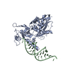 4ngbC 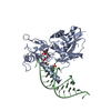 4ngdSC  4ngfC 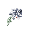 4nggC  4nh3C 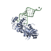 4nh5C  4nh6C 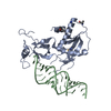 4nhaC C: citing same article ( S: Starting model for refinement |
|---|---|
| Similar structure data |


-Links
 PDBj
PDBj


- Assembly
| Deposited unit | 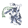
| ||||||||
|---|---|---|---|---|---|---|---|---|---|
| 1 | 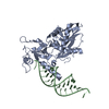
| ||||||||
| 2 | 
| ||||||||
| Unit cell |
|
-Components
| #1: Protein | Mass: 34928.336 Da / Num. of mol.: 1 Fragment: platform-PAZ-connector helix cassette (UNP residues 765-1065) Mutation: K822A/K823A Source method: isolated from a genetically manipulated source Source: (gene. exp.) Homo sapiens (human) / Gene: DICER, DICER1, HERNA, KIAA0928 / Plasmid: pET28b / Production host:  References: UniProt: Q9UPY3, Hydrolases; Acting on ester bonds; Endoribonucleases producing 5'-phosphomonoesters |
|---|---|
| #2: RNA chain | Mass: 3812.320 Da / Num. of mol.: 1 / Source method: obtained synthetically / Details: siRNA |
| #3: Water | ChemComp-HOH /  Mass: 18.015 Da / Num. of mol.: 227 / Source method: isolated from a natural source / Formula: H2O Mass: 18.015 Da / Num. of mol.: 227 / Source method: isolated from a natural source / Formula: H2O |
-Experimental details
-Experiment
| Experiment | Method: X-RAY DIFFRACTION / Number of used crystals: 1 |
|---|
- Sample preparation
| Crystal | Density Matthews: 2.81 Å3/Da / Density % sol: 56.25 % |
|---|---|
| Crystal grow | Temperature: 293 K / Method: vapor diffusion, hanging drop / pH: 7 Details: 0.2 M sodium/potassium tartrate, 0.1 M Bis-Tris propane, pH 7.0, 18-22% PEG3350, VAPOR DIFFUSION, HANGING DROP, temperature 293K |
-Data collection
| Diffraction | Mean temperature: 100 K | |||||||||||||||||||||||||||||||||||||||||||||||||||||||||||||||||||||||||||||
|---|---|---|---|---|---|---|---|---|---|---|---|---|---|---|---|---|---|---|---|---|---|---|---|---|---|---|---|---|---|---|---|---|---|---|---|---|---|---|---|---|---|---|---|---|---|---|---|---|---|---|---|---|---|---|---|---|---|---|---|---|---|---|---|---|---|---|---|---|---|---|---|---|---|---|---|---|---|---|
| Diffraction source | Source: SYNCHROTRON / Site: APS  / Beamline: 24-ID-E / Wavelength: 0.97918 Å / Beamline: 24-ID-E / Wavelength: 0.97918 Å | |||||||||||||||||||||||||||||||||||||||||||||||||||||||||||||||||||||||||||||
| Detector | Type: ADSC QUANTUM 315 / Detector: CCD / Date: Jun 16, 2009 | |||||||||||||||||||||||||||||||||||||||||||||||||||||||||||||||||||||||||||||
| Radiation | Monochromator: Si(220) / Protocol: SINGLE WAVELENGTH / Monochromatic (M) / Laue (L): M / Scattering type: x-ray | |||||||||||||||||||||||||||||||||||||||||||||||||||||||||||||||||||||||||||||
| Radiation wavelength | Wavelength: 0.97918 Å / Relative weight: 1 | |||||||||||||||||||||||||||||||||||||||||||||||||||||||||||||||||||||||||||||
| Reflection | Resolution: 2.1→50 Å / Num. obs: 23726 / % possible obs: 95.2 % / Redundancy: 4.9 % / Biso Wilson estimate: 29.86 Å2 / Rmerge(I) obs: 0.065 / Χ2: 1.056 / Net I/σ(I): 9.4 | |||||||||||||||||||||||||||||||||||||||||||||||||||||||||||||||||||||||||||||
| Reflection shell |
|
-Phasing
| Phasing | Method: molecular replacement |
|---|
- Processing
| Software |
| ||||||||||||||||||||||||||||||||||||||||||||||||||||||||||||||||||||||
|---|---|---|---|---|---|---|---|---|---|---|---|---|---|---|---|---|---|---|---|---|---|---|---|---|---|---|---|---|---|---|---|---|---|---|---|---|---|---|---|---|---|---|---|---|---|---|---|---|---|---|---|---|---|---|---|---|---|---|---|---|---|---|---|---|---|---|---|---|---|---|---|
| Refinement | Method to determine structure: MOLECULAR REPLACEMENT Starting model: PDB ENTRY 4NGD Resolution: 2.104→34.179 Å / Occupancy max: 1 / Occupancy min: 0.37 / FOM work R set: 0.8558 / SU ML: 0.22 / Cross valid method: THROUGHOUT / σ(F): 1.38 / Phase error: 21.82 / Stereochemistry target values: ML
| ||||||||||||||||||||||||||||||||||||||||||||||||||||||||||||||||||||||
| Solvent computation | Shrinkage radii: 0.9 Å / VDW probe radii: 1.11 Å / Solvent model: FLAT BULK SOLVENT MODEL | ||||||||||||||||||||||||||||||||||||||||||||||||||||||||||||||||||||||
| Displacement parameters | Biso max: 85.22 Å2 / Biso mean: 33.3809 Å2 / Biso min: 11.61 Å2 | ||||||||||||||||||||||||||||||||||||||||||||||||||||||||||||||||||||||
| Refinement step | Cycle: LAST / Resolution: 2.104→34.179 Å
| ||||||||||||||||||||||||||||||||||||||||||||||||||||||||||||||||||||||
| Refine LS restraints |
| ||||||||||||||||||||||||||||||||||||||||||||||||||||||||||||||||||||||
| LS refinement shell | Refine-ID: X-RAY DIFFRACTION / Total num. of bins used: 9
| ||||||||||||||||||||||||||||||||||||||||||||||||||||||||||||||||||||||
| Refinement TLS params. | Method: refined / Origin x: 35.038 Å / Origin y: 55.468 Å / Origin z: 6.6969 Å
| ||||||||||||||||||||||||||||||||||||||||||||||||||||||||||||||||||||||
| Refinement TLS group |
|
