 Movie
Movie Controller

[English] 日本語
 Yorodumi
- PDB-4nbj: D-aminoacyl-tRNA deacylase (DTD) from Plasmodium falciparum in co... -
+ Open data

- Basic information
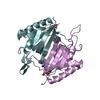
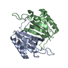
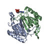
| Entry | Database: PDB / ID: 4nbj | ||||||
|---|---|---|---|---|---|---|---|
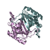
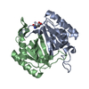
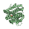
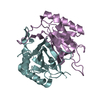
| Title | D-aminoacyl-tRNA deacylase (DTD) from Plasmodium falciparum in complex with D-tyrosyl-3'-aminoadenosine at 2.20 Angstrom resolution | ||||||
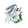
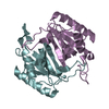
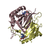
 Components Components | D-tyrosyl-tRNA(Tyr) deacylase | ||||||
 Keywords Keywords | HYDROLASE / DTD / DEACYLASE / DTD-like | ||||||
| Function / homology |  Function and homology information Function and homology informationD-tyrosyl-tRNA(Tyr) deacylase activity / Gly-tRNA(Ala) deacylase activity / D-aminoacyl-tRNA deacylase / tRNA metabolic process / tRNA binding / nucleotide binding / cytoplasm Similarity search - Function | ||||||
| Biological species |  | ||||||
| Method |  X-RAY DIFFRACTION / MOLECULAR REPLACEMENT / Resolution: 2.2 Å X-RAY DIFFRACTION / MOLECULAR REPLACEMENT / Resolution: 2.2 Å | ||||||
 Authors Authors | Ahmad, S. / Routh, S.B. / Kamarthapu, V. / Sankaranarayanan, R. | ||||||
 Citation Citation | Journal: Elife / Year: 2013 Title: Mechanism of chiral proofreading during translation of the genetic code. Authors: Ahmad, S. / Routh, S.B. / Kamarthapu, V. / Chalissery, J. / Muthukumar, S. / Hussain, T. / Kruparani, S.P. / Deshmukh, M.V. / Sankaranarayanan, R. | ||||||
| History |
|
- Structure visualization
| Structure viewer | Molecule: MolmilJmol/JSmol |
|---|
- Downloads & links
-Download
| PDBx/mmCIF format | 4nbj.cif.gz | 269 KB | Display | PDBx/mmCIF format |
|---|---|---|---|---|
| PDB format | pdb4nbj.ent.gz | 220 KB | Display | PDB format |
| PDBx/mmJSON format | 4nbj.json.gz | Tree view | PDBx/mmJSON format | |
| Others |  Other downloads Other downloads |
-Validation report
| Arichive directory | https://data.pdbj.org/pub/pdb/validation_reports/nb/4nbjftp://data.pdbj.org/pub/pdb/validation_reports/nb/4nbj | HTTPS FTP |
|---|
-Related structure data
| Related structure data | 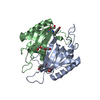 4nbiC  3knfS C: citing same article ( S: Starting model for refinement |
|---|---|
| Similar structure data |
-Links
 PDBj
PDBj- Assembly
| Deposited unit | 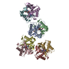
| ||||||||
|---|---|---|---|---|---|---|---|---|---|
| 1 | 
| ||||||||
| 2 | 
| ||||||||
| 3 | 
| ||||||||
| 4 | 
| ||||||||
| Unit cell |
|
-Components
| #1: Protein | Mass: 19233.084 Da / Num. of mol.: 8 Source method: isolated from a genetically manipulated source Source: (gene. exp.) Strain: 3D7 / Gene: DTD, PF11_0095 / Plasmid: pET-21b / Production host:  References: UniProt: Q8IIS0, Hydrolases; Acting on ester bonds #2: Chemical | ChemComp-D3Y /   Mass: 429.430 Da / Num. of mol.: 8 / Source method: obtained synthetically / Formula: C19H23N7O5 Mass: 429.430 Da / Num. of mol.: 8 / Source method: obtained synthetically / Formula: C19H23N7O5#3: Water | ChemComp-HOH / |  Mass: 18.015 Da / Num. of mol.: 375 / Source method: isolated from a natural source / Formula: H2O Mass: 18.015 Da / Num. of mol.: 375 / Source method: isolated from a natural source / Formula: H2O |
|---|
-Experimental details
-Experiment
| Experiment | Method: X-RAY DIFFRACTION / Number of used crystals: 1 |
|---|
- Sample preparation
| Crystal | Density Matthews: 2.24 Å3/Da / Density % sol: 45.06 % / Mosaicity: 1.551 ° |
|---|---|
| Crystal grow | Temperature: 293 K / Method: vapor diffusion, hanging drop / pH: 6 Details: 28% PEG 3350, 0.4M sodium chloride, 0.1M BisTris, pH 6.0, VAPOR DIFFUSION, HANGING DROP, temperature 293K |
-Data collection
| Diffraction | Mean temperature: 93 K | |||||||||||||||||||||||||||||||||||||||||||||||||||||||||||||||||||||||||||||
|---|---|---|---|---|---|---|---|---|---|---|---|---|---|---|---|---|---|---|---|---|---|---|---|---|---|---|---|---|---|---|---|---|---|---|---|---|---|---|---|---|---|---|---|---|---|---|---|---|---|---|---|---|---|---|---|---|---|---|---|---|---|---|---|---|---|---|---|---|---|---|---|---|---|---|---|---|---|---|
| Diffraction source | Source: ROTATING ANODE / Type: RIGAKU FR-E+ SUPERBRIGHT / Wavelength: 1.5418 Å | |||||||||||||||||||||||||||||||||||||||||||||||||||||||||||||||||||||||||||||
| Detector | Type: RIGAKU RAXIS IV++ / Detector: IMAGE PLATE / Date: Apr 16, 2011 / Details: mirrors | |||||||||||||||||||||||||||||||||||||||||||||||||||||||||||||||||||||||||||||
| Radiation | Protocol: SINGLE WAVELENGTH / Monochromatic (M) / Laue (L): M / Scattering type: x-ray | |||||||||||||||||||||||||||||||||||||||||||||||||||||||||||||||||||||||||||||
| Radiation wavelength | Wavelength: 1.5418 Å / Relative weight: 1 | |||||||||||||||||||||||||||||||||||||||||||||||||||||||||||||||||||||||||||||
| Reflection | Resolution: 2.2→25 Å / Num. obs: 69275 / % possible obs: 100 % / Redundancy: 6.5 % / Rmerge(I) obs: 0.118 / Χ2: 1.601 / Net I/σ(I): 23 | |||||||||||||||||||||||||||||||||||||||||||||||||||||||||||||||||||||||||||||
| Reflection shell |
|
- Processing
| Software |
| |||||||||||||||||||||||||||||||||||||||||||||
|---|---|---|---|---|---|---|---|---|---|---|---|---|---|---|---|---|---|---|---|---|---|---|---|---|---|---|---|---|---|---|---|---|---|---|---|---|---|---|---|---|---|---|---|---|---|---|
| Refinement | Method to determine structure: MOLECULAR REPLACEMENT Starting model: 3KNF Resolution: 2.2→25 Å / Cor.coef. Fo:Fc: 0.962 / Cor.coef. Fo:Fc free: 0.934 / Occupancy max: 1 / Occupancy min: 1 / SU B: 7.357 / SU ML: 0.18 / Cross valid method: THROUGHOUT / σ(F): 0 / ESU R: 0.298 / ESU R Free: 0.229 / Stereochemistry target values: MAXIMUM LIKELIHOOD Details: HYDROGENS HAVE BEEN USED IF PRESENT IN THE INPUT U VALUES
| |||||||||||||||||||||||||||||||||||||||||||||
| Solvent computation | Ion probe radii: 0.8 Å / Shrinkage radii: 0.8 Å / VDW probe radii: 1.2 Å / Solvent model: MASK | |||||||||||||||||||||||||||||||||||||||||||||
| Displacement parameters | Biso max: 113.53 Å2 / Biso mean: 48.106 Å2 / Biso min: 24.03 Å2
| |||||||||||||||||||||||||||||||||||||||||||||
| Refinement step | Cycle: LAST / Resolution: 2.2→25 Å
| |||||||||||||||||||||||||||||||||||||||||||||
| Refine LS restraints |
| |||||||||||||||||||||||||||||||||||||||||||||
| LS refinement shell | Resolution: 2.196→2.253 Å / Total num. of bins used: 20
|
