| Entry | Database: PDB / ID: 4n8w
|
|---|
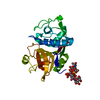
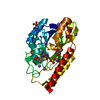
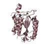
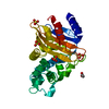
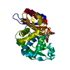
| Title | cathepsin K - chondroitin sulfate complex |
|---|
 Components Components | Cathepsin K |
|---|
 Keywords Keywords | HYDROLASE / collaginase / Glycosaminoglycan |
|---|
| Function / homology |  Function and homology information Function and homology information
cathepsin K / negative regulation of cartilage development / RUNX1 regulates transcription of genes involved in differentiation of keratinocytes / endolysosome lumen / thyroid hormone generation / Trafficking and processing of endosomal TLR / proteoglycan binding / Activation of Matrix Metalloproteinases / Collagen degradation / collagen catabolic process ...cathepsin K / negative regulation of cartilage development / RUNX1 regulates transcription of genes involved in differentiation of keratinocytes / endolysosome lumen / thyroid hormone generation / Trafficking and processing of endosomal TLR / proteoglycan binding / Activation of Matrix Metalloproteinases / Collagen degradation / collagen catabolic process / fibronectin binding / extracellular matrix disassembly / bone resorption / mitophagy / collagen binding / Degradation of the extracellular matrix / cysteine-type peptidase activity / MHC class II antigen presentation / lysosomal lumen / : / lysosome / apical plasma membrane / serine-type endopeptidase activity / external side of plasma membrane / cysteine-type endopeptidase activity / proteolysis / : / extracellular region / nucleoplasmSimilarity search - Function Cathepsin propeptide inhibitor domain (I29) / Cathepsin propeptide inhibitor domain (I29) / Cathepsin propeptide inhibitor domain (I29) / Papain-like cysteine endopeptidase / Cysteine peptidase, asparagine active site / Eukaryotic thiol (cysteine) proteases asparagine active site. / Cysteine peptidase, histidine active site / Eukaryotic thiol (cysteine) proteases histidine active site. / : / Peptidase C1A, papain C-terminal ...Cathepsin propeptide inhibitor domain (I29) / Cathepsin propeptide inhibitor domain (I29) / Cathepsin propeptide inhibitor domain (I29) / Papain-like cysteine endopeptidase / Cysteine peptidase, asparagine active site / Eukaryotic thiol (cysteine) proteases asparagine active site. / Cysteine peptidase, histidine active site / Eukaryotic thiol (cysteine) proteases histidine active site. / : / Peptidase C1A, papain C-terminal / Papain family cysteine protease / Papain family cysteine protease / Cysteine proteinases / Cysteine peptidase, cysteine active site / Eukaryotic thiol (cysteine) proteases cysteine active site. / Cathepsin B; Chain A / Papain-like cysteine peptidase superfamily / Alpha-Beta Complex / Alpha BetaSimilarity search - Domain/homology |
|---|
| Biological species |  Homo sapiens (human) Homo sapiens (human) |
|---|
| Method |  X-RAY DIFFRACTION / SYNCHROTRON / MOLECULAR REPLACEMENT / Resolution: 2.02 Å X-RAY DIFFRACTION / SYNCHROTRON / MOLECULAR REPLACEMENT / Resolution: 2.02 Å |
|---|
 Authors Authors | Aguda, A.H. / Nguyen, N.T. / Bromme, D. / Brayer, G.D. |
|---|
 Citation Citation | Journal: Proc.Natl.Acad.Sci.USA / Year: 2014
Title: Structural basis of collagen fiber degradation by cathepsin K.
Authors: Aguda, A.H. / Panwar, P. / Du, X. / Nguyen, N.T. / Brayer, G.D. / Bromme, D. |
|---|
| History | | Deposition | Oct 18, 2013 | Deposition site: RCSB / Processing site: RCSB |
|---|
| Revision 1.0 | Nov 26, 2014 | Provider: repository / Type: Initial release |
|---|
| Revision 1.1 | Dec 24, 2014 | Group: Database references |
|---|
| Revision 2.0 | Jul 29, 2020 | Group: Atomic model / Data collection ...Atomic model / Data collection / Derived calculations / Structure summary
Category: atom_site / chem_comp ...atom_site / chem_comp / entity / pdbx_branch_scheme / pdbx_chem_comp_identifier / pdbx_entity_branch / pdbx_entity_branch_descriptor / pdbx_entity_branch_link / pdbx_entity_branch_list / pdbx_entity_nonpoly / pdbx_nonpoly_scheme / pdbx_struct_assembly_gen / struct_asym / struct_conn / struct_site / struct_site_gen
Item: _atom_site.B_iso_or_equiv / _atom_site.Cartn_x ..._atom_site.B_iso_or_equiv / _atom_site.Cartn_x / _atom_site.Cartn_y / _atom_site.Cartn_z / _atom_site.auth_asym_id / _atom_site.auth_atom_id / _atom_site.auth_comp_id / _atom_site.auth_seq_id / _atom_site.label_asym_id / _atom_site.label_atom_id / _atom_site.label_comp_id / _atom_site.label_entity_id / _atom_site.occupancy / _atom_site.pdbx_formal_charge / _atom_site.type_symbol / _chem_comp.mon_nstd_flag / _chem_comp.name / _chem_comp.type / _pdbx_struct_assembly_gen.asym_id_list / _struct_conn.pdbx_dist_value / _struct_conn.pdbx_leaving_atom_flag / _struct_conn.pdbx_value_order / _struct_conn.ptnr1_auth_asym_id / _struct_conn.ptnr1_auth_seq_id / _struct_conn.ptnr1_label_asym_id / _struct_conn.ptnr1_label_atom_id / _struct_conn.ptnr2_auth_asym_id / _struct_conn.ptnr2_auth_seq_id / _struct_conn.ptnr2_label_asym_id / _struct_conn.ptnr2_label_atom_id
Description: Carbohydrate remediation / Provider: repository / Type: Remediation |
|---|
| Revision 2.1 | Nov 6, 2024 | Group: Data collection / Database references / Structure summary
Category: chem_comp / chem_comp_atom ...chem_comp / chem_comp_atom / chem_comp_bond / database_2 / pdbx_entry_details / pdbx_modification_feature
Item: _chem_comp.pdbx_synonyms / _database_2.pdbx_DOI ..._chem_comp.pdbx_synonyms / _database_2.pdbx_DOI / _database_2.pdbx_database_accession / _pdbx_entry_details.has_protein_modification |
|---|
|
|---|
 Movie
Movie Controller


 Open data
 Basic information
 Structure visualization
 Downloads & links
 Other downloads


 PDBj
PDBj


 Assembly
 Pichia pastoris (fungus) / References: UniProt: P43235, cathepsin K
 Mass: 18.015 Da / Num. of mol.: 79 / Source method: isolated from a natural source / Formula: H2O
 Sample preparation
 / Beamline: BL7-1 / Wavelength: 0.97 Å
 Processing