 Movie
Movie Controller

[English] 日本語
 Yorodumi
- PDB-4mo9: Crystal Structure of TroA-like Periplasmic Binding Protein FepB f... -
+ Open data

- Basic information
| Entry | Database: PDB / ID: 4mo9 | ||||||
|---|---|---|---|---|---|---|---|
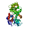
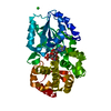
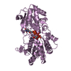
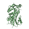
| Title | Crystal Structure of TroA-like Periplasmic Binding Protein FepB from Veillonella parvula | ||||||
 Components Components | Periplasmic binding protein | ||||||
 Keywords Keywords | SOLUTE-BINDING PROTEIN / Structural Genomics / PSI-Biology / Midwest Center for Structural Genomics / MCSG / periplasmic binding protein / solute binding protein-fold / PROTEIN BINDING | ||||||
| Function / homology | ABC transporter periplasmic binding domain / Periplasmic binding protein / Iron siderophore/cobalamin periplasmic-binding domain profile. / trimethylamine oxide / Periplasmic binding protein Function and homology information Function and homology information | ||||||
| Biological species |  Veillonella parvula (bacteria) Veillonella parvula (bacteria) | ||||||
| Method |  X-RAY DIFFRACTION / SYNCHROTRON / SAD / Resolution: 1.925 Å X-RAY DIFFRACTION / SYNCHROTRON / SAD / Resolution: 1.925 Å | ||||||
 Authors Authors | Kim, Y. / Wu, R. / Endres, M. / Joachimiak, A. / Midwest Center for Structural Genomics (MCSG) | ||||||
 Citation Citation | Journal: To be Published Title: Crystal Structure of TroA-like Periplasmic Binding Protein FepB from Veillonella parvula Authors: Kim, Y. / Wu, R. / Endres, M. / Joachimiak, A. / Midwest Center for Structural Genomics (MCSG) | ||||||
| History |
|
- Structure visualization
| Structure viewer | Molecule: MolmilJmol/JSmol |
|---|
- Downloads & links
-Download
| PDBx/mmCIF format | 4mo9.cif.gz | 155.5 KB | Display | PDBx/mmCIF format |
|---|---|---|---|---|
| PDB format | pdb4mo9.ent.gz | 123.2 KB | Display | PDB format |
| PDBx/mmJSON format | 4mo9.json.gz | Tree view | PDBx/mmJSON format | |
| Others |  Other downloads Other downloads |
-Validation report
| Arichive directory | https://data.pdbj.org/pub/pdb/validation_reports/mo/4mo9ftp://data.pdbj.org/pub/pdb/validation_reports/mo/4mo9 | HTTPS FTP |
|---|
-Related structure data
| Similar structure data | |
|---|---|
| Other databases |


-Links
 PDBj
PDBj- Assembly
| Deposited unit | 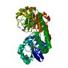
| ||||||||
|---|---|---|---|---|---|---|---|---|---|
| 1 |
| ||||||||
| Unit cell |
|
-Components
| #1: Protein | Mass: 41671.336 Da / Num. of mol.: 1 / Fragment: UNP residues 25-390 Source method: isolated from a genetically manipulated source Source: (gene. exp.) Veillonella parvula (bacteria) / Strain: DSM 2008 / Gene: Vpar_0195 / Plasmid: pMCSG68 / Production host: |
|---|---|
| #2: Chemical | ChemComp-TMO /   Mass: 75.110 Da / Num. of mol.: 1 / Source method: obtained synthetically / Formula: C3H9NO Mass: 75.110 Da / Num. of mol.: 1 / Source method: obtained synthetically / Formula: C3H9NO |
| #3: Chemical | ChemComp-GOL /   Mass: 92.094 Da / Num. of mol.: 1 / Source method: obtained synthetically / Formula: C3H8O3 Mass: 92.094 Da / Num. of mol.: 1 / Source method: obtained synthetically / Formula: C3H8O3 |
| #4: Water | ChemComp-HOH /  Mass: 18.015 Da / Num. of mol.: 198 / Source method: isolated from a natural source / Formula: H2O Mass: 18.015 Da / Num. of mol.: 198 / Source method: isolated from a natural source / Formula: H2O |
| Has protein modification | Y |
-Experimental details
-Experiment
| Experiment | Method: X-RAY DIFFRACTION / Number of used crystals: 1 |
|---|
- Sample preparation
| Crystal | Density Matthews: 2.25 Å3/Da / Density % sol: 45.31 % |
|---|---|
| Crystal grow | Temperature: 289 K / Method: vapor diffusion, sitting drop / pH: 8.5 Details: 0.2 M Trimethylamine N-oxide, 0.1 M Tris pH 8.5, 20 %(w/v) PEGMME2000, VAPOR DIFFUSION, SITTING DROP, temperature 289K |
-Data collection
| Diffraction | Mean temperature: 100 K |
|---|---|
| Diffraction source | Source: SYNCHROTRON / Site: APS  / Beamline: 19-ID / Wavelength: 0.97987 Å / Beamline: 19-ID / Wavelength: 0.97987 Å |
| Detector | Type: ADSC QUANTUM 315r / Detector: CCD / Date: Apr 22, 2013 / Details: mirrors |
| Radiation | Monochromator: double crystal monochromator / Protocol: SINGLE WAVELENGTH / Monochromatic (M) / Laue (L): M / Scattering type: x-ray |
| Radiation wavelength | Wavelength: 0.97987 Å / Relative weight: 1 |
| Reflection | Resolution: 1.93→50 Å / Num. all: 29285 / Num. obs: 29285 / % possible obs: 99.9 % / Observed criterion σ(F): 0 / Observed criterion σ(I): 0 / Redundancy: 5.8 % / Biso Wilson estimate: 32.75 Å2 / Rsym value: 0.103 / Net I/σ(I): 9.9 |
| Reflection shell | Resolution: 1.93→1.96 Å / Redundancy: 4.6 % / Mean I/σ(I) obs: 2 / Num. unique all: 1450 / Rsym value: 0.792 / % possible all: 100 |
- Processing
| Software |
| ||||||||||||||||||||||||||||||||||||||||||||||||||||||||||||||||||||||||||||||||||||||||||||||||||||
|---|---|---|---|---|---|---|---|---|---|---|---|---|---|---|---|---|---|---|---|---|---|---|---|---|---|---|---|---|---|---|---|---|---|---|---|---|---|---|---|---|---|---|---|---|---|---|---|---|---|---|---|---|---|---|---|---|---|---|---|---|---|---|---|---|---|---|---|---|---|---|---|---|---|---|---|---|---|---|---|---|---|---|---|---|---|---|---|---|---|---|---|---|---|---|---|---|---|---|---|---|---|
| Refinement | Method to determine structure: SAD / Resolution: 1.925→36.361 Å / SU ML: 0.19 / Isotropic thermal model: mixed / Cross valid method: THROUGHOUT / σ(F): 0 / Phase error: 21.49 / Stereochemistry target values: ML
| ||||||||||||||||||||||||||||||||||||||||||||||||||||||||||||||||||||||||||||||||||||||||||||||||||||
| Solvent computation | Shrinkage radii: 0.9 Å / VDW probe radii: 1.11 Å / Solvent model: FLAT BULK SOLVENT MODEL | ||||||||||||||||||||||||||||||||||||||||||||||||||||||||||||||||||||||||||||||||||||||||||||||||||||
| Displacement parameters | Biso mean: 40.5 Å2 | ||||||||||||||||||||||||||||||||||||||||||||||||||||||||||||||||||||||||||||||||||||||||||||||||||||
| Refinement step | Cycle: LAST / Resolution: 1.925→36.361 Å
| ||||||||||||||||||||||||||||||||||||||||||||||||||||||||||||||||||||||||||||||||||||||||||||||||||||
| Refine LS restraints |
| ||||||||||||||||||||||||||||||||||||||||||||||||||||||||||||||||||||||||||||||||||||||||||||||||||||
| LS refinement shell | Refine-ID: X-RAY DIFFRACTION
| ||||||||||||||||||||||||||||||||||||||||||||||||||||||||||||||||||||||||||||||||||||||||||||||||||||
| Refinement TLS params. | Method: refined / Refine-ID: X-RAY DIFFRACTION
| ||||||||||||||||||||||||||||||||||||||||||||||||||||||||||||||||||||||||||||||||||||||||||||||||||||
| Refinement TLS group |
|
