 Movie
Movie Controller

[English] 日本語
 Yorodumi
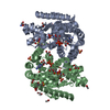
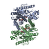
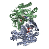
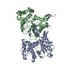
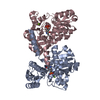
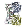
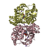
- PDB-4mdh: REFINED CRYSTAL STRUCTURE OF CYTOPLASMIC MALATE DEHYDROGENASE AT ... -
+ Open data

- Basic information
| Entry | Database: PDB / ID: 4mdh | |||||||||
|---|---|---|---|---|---|---|---|---|---|---|
| Title | REFINED CRYSTAL STRUCTURE OF CYTOPLASMIC MALATE DEHYDROGENASE AT 2.5-ANGSTROMS RESOLUTION | |||||||||
 Components Components | CYTOPLASMIC MALATE DEHYDROGENASE | |||||||||
 Keywords Keywords | OXIDOREDUCTASE(NAD(A)-CHOH(D)) | |||||||||
| Function / homology |  Function and homology information Function and homology information(2R)-hydroxyphenylpyruvate reductase [NAD(P)H] activity / Malate-aspartate shuttle / (S)-malate dehydrogenase (NAD+, oxaloacetate-forming) / L-malate dehydrogenase (NAD+) activity / malate metabolic process / oxaloacetate metabolic process / NAD+ metabolic process / tricarboxylic acid cycle / NAD binding / cytosol Similarity search - Function | |||||||||
| Biological species |  | |||||||||
| Method |  X-RAY DIFFRACTION / Resolution: 2.5 Å X-RAY DIFFRACTION / Resolution: 2.5 Å | |||||||||
 Authors Authors | Birktoft, J.J. / Banaszak, L.J. | |||||||||
 Citation Citation | Journal: Biochemistry / Year: 1989 Title: Refined crystal structure of cytoplasmic malate dehydrogenase at 2.5-A resolution. Authors: Birktoft, J.J. / Rhodes, G. / Banaszak, L.J. #1: Journal: To be PublishedTitle: Comparison of the Molecular Structures of Cytoplasmic and Mitochondrial Malate Dehydrogenase Authors: Birktoft, J.J. / Fu, Z. / Carnahan, G.E. / Rhodes, G. / Roderick, S.L. / Banaszak, L.J. #2: Journal: Biochemistry / Year: 1987Title: Structure of Porcine Heart Cytoplasmic Malate Dehydrogenase. Combining X-Ray Diffraction and Chemical Sequence Data in Structural Studies Authors: Birktoft, J.J. / Bradshaw, R.A. / Banaszak, L.J. #3: Journal: J.Biol.Chem. / Year: 1983Title: The Presence of a Histidine-Aspartic Acid Pair in the Active Site of 2-Hydroxyacid Dehydrogenases. X-Ray Refinement of Cytoplasmic Malate Dehydrogenase Authors: Birktoft, J.J. / Banaszak, L.J. #4: Journal: Molecular Structure and Biological Activity / Year: 1982Title: The Interactions of Nad/Nadh with 2-Hydroxy Acid Dehydrogenases Authors: Birktoft, J.J. / Fernley, R.T. / Bradshaw, R.A. / Banaszak, L.J. #5: Journal: Proc.Natl.Acad.Sci.USA / Year: 1982Title: Amino Acid Sequence Homology Among the 2-Hydroxy Acid Dehydrogenases. Mitochondrial and Cytoplasmic Malate Dehydrogenases Form a Homologous System with Lactate Dehydrogenase Authors: Birktoft, J.J. / Fernley, R.T. / Bradshaw, R.A. / Banaszak, L.J. #6: Journal: STRUCTURE AND CONFORMATION OF NUCLEIC ACIDS AND PROTEIN-NUCLEIC ACID INTERACTIONS : PROCEEDINGS OF THE FOURTH ANNUAL HARRY STEENBOCK SYMPOSIUM, JUNE 16-19, 1974, MADISON, WISCONSINYear: 1975 Title: Nicotinamide Adenine Dinucleotide and the Active Site of Cytoplasmic Malate Dehydrogenase Authors: Banaszak, L.J. / Webb, L.E. #7: Journal: Biochemistry / Year: 1973Title: Conformation of Nicotinamide Adenine Dinucleotide Bound to Cytoplasmic Malate Dehydrogenase Authors: Webb, L.E. / Hill, E.J. / Banaszak, L.J. #8: Journal: J.Mol.Biol. / Year: 1972Title: Polypeptide Conformation of Cytoplasmic Malate Dehydrogenase from an Electron Density Map at 3.0 Angstroms Resolution Authors: Hill, E. / Tsernoglou, D. / Webb, L. / Banaszak, L.J. #9: Journal: Biochem.Biophys.Res.Commun. / Year: 1972Title: The Identification of an Asymmetric Complex of Nicotinamide Adenine Dinucleotide and Pig Heart Cytoplasmic Malate Dehydrogenase Authors: Glatthaar, B.E. / Banaszak, L.J. / Bradshaw, R.A. #10: Journal: J.Mol.Biol. / Year: 1972Title: Cytoplasmic Malate Dehydrogenase-Heavy Atom Derivatives and Low Resolution Structure Authors: Tsernoglou, D. / Hill, E. / Banaszak, L.J. #11: Journal: Cold Spring Harbor Symp.Quant.Biol. / Year: 1972Title: Structural Studies on Heart Muscle Malate Dehydrogenases Authors: Tsernoglou, D. / Hill, E. / Banaszak, L.J. | |||||||||
| History |
| |||||||||
| Remark 700 | SHEET IN SHEET S2 THE REGULAR HYDROGEN BONDING PATTERN IS DISRUPTED BY A THREE RESIDUE BULGE ...SHEET IN SHEET S2 THE REGULAR HYDROGEN BONDING PATTERN IS DISRUPTED BY A THREE RESIDUE BULGE (RESIDUES 194 -196) IN STRAND 2. IN SHEET S1 A BETA BULGE IS FOUND IN STRAND 1 AT RESIDUE 64. |
- Structure visualization
| Structure viewer | Molecule: MolmilJmol/JSmol |
|---|
- Downloads & links
-Download
| PDBx/mmCIF format | 4mdh.cif.gz | 152.1 KB | Display | PDBx/mmCIF format |
|---|---|---|---|---|
| PDB format | pdb4mdh.ent.gz | 120.3 KB | Display | PDB format |
| PDBx/mmJSON format | 4mdh.json.gz | Tree view | PDBx/mmJSON format | |
| Others |  Other downloads Other downloads |
-Validation report
| Arichive directory | https://data.pdbj.org/pub/pdb/validation_reports/md/4mdhftp://data.pdbj.org/pub/pdb/validation_reports/md/4mdh | HTTPS FTP |
|---|
-Related structure data
| Similar structure data |
|---|

-Links
 PDBj
PDBj


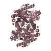
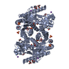
- Assembly
| Deposited unit | 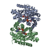
| ||||||||
|---|---|---|---|---|---|---|---|---|---|
| 1 |
| ||||||||
| Unit cell |
| ||||||||
| Atom site foot note | 1: RESIDUES PRO A 131 AND PRO B 131 ARE CIS PROLINES. / 2: SEE REMARK 5. | ||||||||
| Noncrystallographic symmetry (NCS) | NCS oper: (Code: given Matrix: (-0.86554, 0.46781, -0.17888), Vector: |
-Components
| #1: Protein | Mass: 36393.980 Da / Num. of mol.: 2 Source method: isolated from a genetically manipulated source Source: (gene. exp.) References: UniProt: P11708, (S)-malate dehydrogenase (NAD+, oxaloacetate-forming) #2: Chemical |   Mass: 96.063 Da / Num. of mol.: 2 / Source method: obtained synthetically / Formula: SO4 Mass: 96.063 Da / Num. of mol.: 2 / Source method: obtained synthetically / Formula: SO4#3: Chemical |   Mass: 663.425 Da / Num. of mol.: 2 / Source method: obtained synthetically / Formula: C21H27N7O14P2 / Comment: NAD*YM Mass: 663.425 Da / Num. of mol.: 2 / Source method: obtained synthetically / Formula: C21H27N7O14P2 / Comment: NAD*YM#4: Water | ChemComp-HOH / |  Mass: 18.015 Da / Num. of mol.: 471 / Source method: isolated from a natural source / Formula: H2O Mass: 18.015 Da / Num. of mol.: 471 / Source method: isolated from a natural source / Formula: H2OHas protein modification | Y | Nonpolymer details | THREE DIFFERENT COMPOUNDS CONTAINING HEAVY ATOMS WERE FOUND TO BIND TO CMDH. EACH ASYMMETRIC UNIT ...THREE DIFFERENT COMPOUNDS CONTAINING | |
|---|
-Experimental details
-Experiment
| Experiment | Method: X-RAY DIFFRACTION |
|---|
- Sample preparation
| Crystal | Density Matthews: 2.43 Å3/Da / Density % sol: 49.46 % |
|---|
-Data collection
| Reflection | *PLUS Highest resolution: 2.5 Å / Lowest resolution: 6 Å / Num. all: 23373 / Num. obs: 22910 |
|---|
- Processing
| Software | Name: TNT / Classification: refinement | ||||||||||||||||||||||||||||||
|---|---|---|---|---|---|---|---|---|---|---|---|---|---|---|---|---|---|---|---|---|---|---|---|---|---|---|---|---|---|---|---|
| Refinement | Resolution: 2.5→6 Å / Rfactor obs: 0.167 | ||||||||||||||||||||||||||||||
| Refine analyze | Luzzati coordinate error obs: 0.22 Å | ||||||||||||||||||||||||||||||
| Refinement step | Cycle: LAST / Resolution: 2.5→6 Å
| ||||||||||||||||||||||||||||||
| Refine LS restraints |
| ||||||||||||||||||||||||||||||
| Refinement | *PLUS Num. reflection obs: 22910 / Rfactor obs: 0.167 | ||||||||||||||||||||||||||||||
| Solvent computation | *PLUS | ||||||||||||||||||||||||||||||
| Displacement parameters | *PLUS | ||||||||||||||||||||||||||||||
| Refine LS restraints | *PLUS
|
