 Movie
Movie Controller

[English] 日本語
 Yorodumi
- PDB-4m05: Crystal Structure of Mutant Chlorite Dismutase from Candidatus Ni... -
+ Open data

- Basic information
| Entry | Database: PDB / ID: 4m05 | ||||||
|---|---|---|---|---|---|---|---|
| Title | Crystal Structure of Mutant Chlorite Dismutase from Candidatus Nitrospira defluvii R173E | ||||||
 Components Components | Chlorite dismutase | ||||||
 Keywords Keywords | OXIDOREDUCTASE / Ferredoxin-like fold | ||||||
| Function / homology |  Function and homology information Function and homology informationchlorite O2-lyase / chlorite O2-lyase activity / heme binding / metal ion binding Similarity search - Function | ||||||
| Biological species |  Candidatus Nitrospira defluvii (bacteria) Candidatus Nitrospira defluvii (bacteria) | ||||||
| Method |  X-RAY DIFFRACTION / SYNCHROTRON / MOLECULAR REPLACEMENT / Resolution: 2.28 Å X-RAY DIFFRACTION / SYNCHROTRON / MOLECULAR REPLACEMENT / Resolution: 2.28 Å | ||||||
 Authors Authors | Gysel, K. / Hagmueller, A. / Djinovic-Carugo, K. | ||||||
 Citation Citation | Journal: Biochemistry / Year: 2014 Title: Manipulating conserved heme cavity residues of chlorite dismutase: effect on structure, redox chemistry, and reactivity. Authors: Hofbauer, S. / Gysel, K. / Bellei, M. / Hagmuller, A. / Schaffner, I. / Mlynek, G. / Kostan, J. / Pirker, K.F. / Daims, H. / Furtmuller, P.G. / Battistuzzi, G. / Djinovic-Carugo, K. / Obinger, C. | ||||||
| History |
|
- Structure visualization
| Structure viewer | Molecule: MolmilJmol/JSmol |
|---|
- Downloads & links
-Download
| PDBx/mmCIF format | 4m05.cif.gz | 504.1 KB | Display | PDBx/mmCIF format |
|---|---|---|---|---|
| PDB format | pdb4m05.ent.gz | 423.5 KB | Display | PDB format |
| PDBx/mmJSON format | 4m05.json.gz | Tree view | PDBx/mmJSON format | |
| Others |  Other downloads Other downloads |
-Validation report
| Arichive directory | https://data.pdbj.org/pub/pdb/validation_reports/m0/4m05ftp://data.pdbj.org/pub/pdb/validation_reports/m0/4m05 | HTTPS FTP |
|---|
-Related structure data


-Links
 PDBj
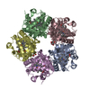
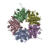
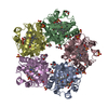
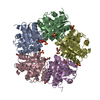
PDBj- Assembly
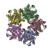
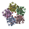
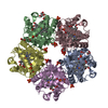
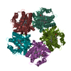
| Deposited unit | 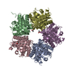
| ||||||||
|---|---|---|---|---|---|---|---|---|---|
| 1 |
| ||||||||
| Unit cell |
| ||||||||
| Noncrystallographic symmetry (NCS) | NCS domain: (Details: chain E and segid E) NCS domain segments: (Selection details: chain 'E' and segid 'E ') |
-Components
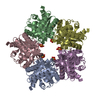
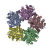
| #1: Protein | Mass: 27451.156 Da / Num. of mol.: 5 / Fragment: UNP residues 27-264 / Mutation: R173E Source method: isolated from a genetically manipulated source Source: (gene. exp.) Candidatus Nitrospira defluvii (bacteria)Gene: cld, cld1, NIDE1387 / Plasmid: pET21b Strep TEV / Production host: #2: Chemical | ChemComp-HEM /   Mass: 616.487 Da / Num. of mol.: 5 / Source method: obtained synthetically / Formula: C34H32FeN4O4 Mass: 616.487 Da / Num. of mol.: 5 / Source method: obtained synthetically / Formula: C34H32FeN4O4#3: Chemical |   Mass: 59.044 Da / Num. of mol.: 2 / Source method: obtained synthetically / Formula: C2H3O2 Mass: 59.044 Da / Num. of mol.: 2 / Source method: obtained synthetically / Formula: C2H3O2#4: Chemical | ChemComp-EDO / |   Mass: 62.068 Da / Num. of mol.: 1 / Source method: obtained synthetically / Formula: C2H6O2 Mass: 62.068 Da / Num. of mol.: 1 / Source method: obtained synthetically / Formula: C2H6O2#5: Water | ChemComp-HOH / |  Mass: 18.015 Da / Num. of mol.: 133 / Source method: isolated from a natural source / Formula: H2O Mass: 18.015 Da / Num. of mol.: 133 / Source method: isolated from a natural source / Formula: H2O |
|---|
-Experimental details
-Experiment
| Experiment | Method: X-RAY DIFFRACTION / Number of used crystals: 3 |
|---|
- Sample preparation
| Crystal | Density Matthews: 2.79 Å3/Da / Density % sol: 55.86 % |
|---|---|
| Crystal grow | Temperature: 277 K / Method: vapor diffusion / pH: 4.5 Details: 40% (v/v) ethylene glycol, 0.1 M NaAc pH 4.5, 3:1, sitting, vapor diffusion, temperature 277K, VAPOR DIFFUSION |
-Data collection
| Diffraction | Mean temperature: 100 K |
|---|---|
| Diffraction source | Source: SYNCHROTRON / Site: ESRF  / Beamline: ID23-1 / Wavelength: 0.976 Å / Beamline: ID23-1 / Wavelength: 0.976 Å |
| Detector | Type: MARMOSAIC 225 mm CCD / Detector: CCD / Date: Apr 17, 2011 |
| Radiation | Monochromator: Si(111) monochromator / Protocol: SINGLE WAVELENGTH / Monochromatic (M) / Laue (L): M / Scattering type: x-ray |
| Radiation wavelength | Wavelength: 0.976 Å / Relative weight: 1 |
| Reflection | Resolution: 2.28→44.22 Å / Num. obs: 72249 / % possible obs: 99.6 % / Redundancy: 9.8 % / Biso Wilson estimate: 30.08 Å2 Details: CC1/2 FOR WHOLE RANGE=0.993, CC1/2 FOR HIGH RESOLUTION SHELL=0.129 Rmerge(I) obs: 0.371 / Net I/σ(I): 5.3 |
| Reflection shell | Resolution: 2.28→2.361 Å / Redundancy: 5.8 % / Rmerge(I) obs: 6.865 / Mean I/σ(I) obs: 0.23 / Num. unique all: 6981 |
- Processing
| Software |
| |||||||||||||||||||||||||||||||||||||||||||||||||||||||||||||||||||||||||||||||||||||||||||||||||||||||||||||||||||||||||||||||||||||||||||||||||||||||||||||||||||||||||||||||||||||||||||||||||||||||||||||||||||||||||
|---|---|---|---|---|---|---|---|---|---|---|---|---|---|---|---|---|---|---|---|---|---|---|---|---|---|---|---|---|---|---|---|---|---|---|---|---|---|---|---|---|---|---|---|---|---|---|---|---|---|---|---|---|---|---|---|---|---|---|---|---|---|---|---|---|---|---|---|---|---|---|---|---|---|---|---|---|---|---|---|---|---|---|---|---|---|---|---|---|---|---|---|---|---|---|---|---|---|---|---|---|---|---|---|---|---|---|---|---|---|---|---|---|---|---|---|---|---|---|---|---|---|---|---|---|---|---|---|---|---|---|---|---|---|---|---|---|---|---|---|---|---|---|---|---|---|---|---|---|---|---|---|---|---|---|---|---|---|---|---|---|---|---|---|---|---|---|---|---|---|---|---|---|---|---|---|---|---|---|---|---|---|---|---|---|---|---|---|---|---|---|---|---|---|---|---|---|---|---|---|---|---|---|---|---|---|---|---|---|---|---|---|---|---|---|---|---|---|---|
| Refinement | Method to determine structure: MOLECULAR REPLACEMENT / Resolution: 2.28→44.22 Å / Occupancy max: 1 / Occupancy min: 0.61 / FOM work R set: 0.6284 / SU ML: 0.6 / σ(F): 1.19 / Phase error: 41.91 / Stereochemistry target values: ML
| |||||||||||||||||||||||||||||||||||||||||||||||||||||||||||||||||||||||||||||||||||||||||||||||||||||||||||||||||||||||||||||||||||||||||||||||||||||||||||||||||||||||||||||||||||||||||||||||||||||||||||||||||||||||||
| Solvent computation | Shrinkage radii: 0.9 Å / VDW probe radii: 1.11 Å / Solvent model: FLAT BULK SOLVENT MODEL | |||||||||||||||||||||||||||||||||||||||||||||||||||||||||||||||||||||||||||||||||||||||||||||||||||||||||||||||||||||||||||||||||||||||||||||||||||||||||||||||||||||||||||||||||||||||||||||||||||||||||||||||||||||||||
| Displacement parameters | Biso max: 207.95 Å2 / Biso mean: 73.3882 Å2 / Biso min: 33.16 Å2 | |||||||||||||||||||||||||||||||||||||||||||||||||||||||||||||||||||||||||||||||||||||||||||||||||||||||||||||||||||||||||||||||||||||||||||||||||||||||||||||||||||||||||||||||||||||||||||||||||||||||||||||||||||||||||
| Refinement step | Cycle: LAST / Resolution: 2.28→44.22 Å
| |||||||||||||||||||||||||||||||||||||||||||||||||||||||||||||||||||||||||||||||||||||||||||||||||||||||||||||||||||||||||||||||||||||||||||||||||||||||||||||||||||||||||||||||||||||||||||||||||||||||||||||||||||||||||
| Refine LS restraints |
| |||||||||||||||||||||||||||||||||||||||||||||||||||||||||||||||||||||||||||||||||||||||||||||||||||||||||||||||||||||||||||||||||||||||||||||||||||||||||||||||||||||||||||||||||||||||||||||||||||||||||||||||||||||||||
| Refine LS restraints NCS | Number: 5801 / Type: POSITIONAL / Rms dev position: 7.878 Å | |||||||||||||||||||||||||||||||||||||||||||||||||||||||||||||||||||||||||||||||||||||||||||||||||||||||||||||||||||||||||||||||||||||||||||||||||||||||||||||||||||||||||||||||||||||||||||||||||||||||||||||||||||||||||
| LS refinement shell | Refine-ID: X-RAY DIFFRACTION / Total num. of bins used: 30
| |||||||||||||||||||||||||||||||||||||||||||||||||||||||||||||||||||||||||||||||||||||||||||||||||||||||||||||||||||||||||||||||||||||||||||||||||||||||||||||||||||||||||||||||||||||||||||||||||||||||||||||||||||||||||
| Refinement TLS params. | Method: refined / Origin x: -8.6181 Å / Origin y: 0.0044 Å / Origin z: -28.8415 Å
| |||||||||||||||||||||||||||||||||||||||||||||||||||||||||||||||||||||||||||||||||||||||||||||||||||||||||||||||||||||||||||||||||||||||||||||||||||||||||||||||||||||||||||||||||||||||||||||||||||||||||||||||||||||||||
| Refinement TLS group |
|
