 Movie
Movie Controller

[English] 日本語
 Yorodumi
- PDB-4ly3: Crystal structure of WlaRD, a sugar 3N-formyl transferase in the ... -
+ Open data

- Basic information
| Entry | Database: PDB / ID: 4ly3 | ||||||
|---|---|---|---|---|---|---|---|
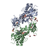
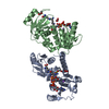
| Title | Crystal structure of WlaRD, a sugar 3N-formyl transferase in the presence of dTPD-Qui3N, dTDP-Qui3NFo, and THF | ||||||
 Components Components | WlaRD a sugar 3N formyltransferase | ||||||
 Keywords Keywords | TRANSFERASE / formyltransferase | ||||||
| Function / homology | Rossmann fold - #12230 / Rossmann fold / 3-Layer(aba) Sandwich / Alpha Beta / : / Chem-1YJ / Chem-T3Q / :  Function and homology information Function and homology information | ||||||
| Biological species |  Campylobacter jejuni subsp. jejuni (Campylobacter) Campylobacter jejuni subsp. jejuni (Campylobacter) | ||||||
| Method |  X-RAY DIFFRACTION / MOLECULAR REPLACEMENT / Resolution: 1.9 Å X-RAY DIFFRACTION / MOLECULAR REPLACEMENT / Resolution: 1.9 Å | ||||||
 Authors Authors | Thoden, J.B. / Goneau, M.-F. / Gilbert, M. / Holden, H.M. | ||||||
 Citation Citation | Journal: Biochemistry / Year: 2013 Title: Structure of a sugar N-formyltransferase from Campylobacter jejuni. Authors: Thoden, J.B. / Goneau, M.F. / Gilbert, M. / Holden, H.M. | ||||||
| History |
|
- Structure visualization
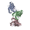
| Structure viewer | Molecule: MolmilJmol/JSmol |
|---|
- Downloads & links
-Download
| PDBx/mmCIF format | 4ly3.cif.gz | 143.4 KB | Display | PDBx/mmCIF format |
|---|---|---|---|---|
| PDB format | pdb4ly3.ent.gz | 110.2 KB | Display | PDB format |
| PDBx/mmJSON format | 4ly3.json.gz | Tree view | PDBx/mmJSON format | |
| Others |  Other downloads Other downloads |
-Validation report
| Summary document | 4ly3_validation.pdf.gz | 1.5 MB | Display | wwPDB validaton report |
|---|---|---|---|---|
| Full document | 4ly3_full_validation.pdf.gz | 1.5 MB | Display | |
| Data in XML | 4ly3_validation.xml.gz | 29.4 KB | Display | |
| Data in CIF | 4ly3_validation.cif.gz | 44.1 KB | Display | |
| Arichive directory | https://data.pdbj.org/pub/pdb/validation_reports/ly/4ly3ftp://data.pdbj.org/pub/pdb/validation_reports/ly/4ly3 | HTTPS FTP |
-Related structure data
| Related structure data | 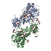 4lxqSC  4lxtC  4lxuC  4lxxC 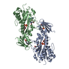 4lxyC 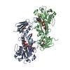 4ly0C S: Starting model for refinement C: citing same article ( |
|---|---|
| Similar structure data |


-Links
 PDBj
PDBj- Assembly
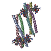
| Deposited unit | 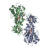
| ||||||||
|---|---|---|---|---|---|---|---|---|---|
| 1 |
| ||||||||
| Unit cell |
|
-Components
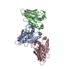
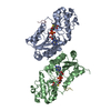
-Protein , 1 types, 2 molecules AB
| #1: Protein | Mass: 31936.607 Da / Num. of mol.: 2 Source method: isolated from a genetically manipulated source Source: (gene. exp.) Campylobacter jejuni subsp. jejuni (Campylobacter)Strain: 81116 / Gene: C8J_1081 / Plasmid: pET28 / Production host: |
|---|
-Non-polymers , 5 types, 615 molecules 
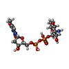

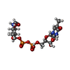

| #2: Chemical |  Mass: 445.429 Da / Num. of mol.: 2 / Source method: obtained synthetically / Formula: C19H23N7O6 Mass: 445.429 Da / Num. of mol.: 2 / Source method: obtained synthetically / Formula: C19H23N7O6#3: Chemical | ChemComp-T3Q / [( |  Mass: 547.345 Da / Num. of mol.: 1 / Source method: obtained synthetically / Formula: C16H27N3O14P2 Mass: 547.345 Da / Num. of mol.: 1 / Source method: obtained synthetically / Formula: C16H27N3O14P2#4: Chemical |  Mass: 209.263 Da / Num. of mol.: 2 / Source method: obtained synthetically / Formula: C7H15NO4S / Comment: pH buffer*YM Mass: 209.263 Da / Num. of mol.: 2 / Source method: obtained synthetically / Formula: C7H15NO4S / Comment: pH buffer*YM#5: Chemical | ChemComp-1YF / [( |  Mass: 575.355 Da / Num. of mol.: 1 / Source method: obtained synthetically / Formula: C17H27N3O15P2 Mass: 575.355 Da / Num. of mol.: 1 / Source method: obtained synthetically / Formula: C17H27N3O15P2#6: Water | ChemComp-HOH / | Mass: 18.015 Da / Num. of mol.: 609 / Source method: isolated from a natural source / Formula: H2O |
|---|
-Experimental details
-Experiment
| Experiment | Method: X-RAY DIFFRACTION / Number of used crystals: 1 |
|---|
- Sample preparation
| Crystal | Density Matthews: 3.12 Å3/Da / Density % sol: 60.53 % |
|---|---|
| Crystal grow | Temperature: 295 K / Method: vapor diffusion, hanging drop / pH: 7 Details: 11-15% PEG 8000, 200 mM NaCl, 5 mM TDP, 5 mM N-5-formyl-THF, pH 7, VAPOR DIFFUSION, HANGING DROP, temperature 295K |
-Data collection
| Diffraction | Mean temperature: 100 K |
|---|---|
| Diffraction source | Source: ROTATING ANODE / Type: RIGAKU RU200 / Wavelength: 1.5418 Å |
| Detector | Type: Bruker Platinum 135 / Detector: CCD / Date: May 20, 2013 / Details: mirrors |
| Radiation | Monochromator: montel optics / Protocol: SINGLE WAVELENGTH / Monochromatic (M) / Laue (L): M / Scattering type: x-ray |
| Radiation wavelength | Wavelength: 1.5418 Å / Relative weight: 1 |
| Reflection | Resolution: 1.9→50 Å / Num. all: 58443 / Num. obs: 58443 / % possible obs: 92.8 % / Observed criterion σ(F): 0 / Observed criterion σ(I): 0 / Redundancy: 3.6 % / Rmerge(I) obs: 0.05 / Rsym value: 0.05 / Net I/σ(I): 15.9 |
- Processing
| Software |
| ||||||||||||||||||||||||||||||||||||||||||||||||||||||||||||||||||||||||||||||||||||||||||||||||||||||||||||||||||||||||||||||||||||||||||||||||||||||||||||||||||||||||||||||||||||||
|---|---|---|---|---|---|---|---|---|---|---|---|---|---|---|---|---|---|---|---|---|---|---|---|---|---|---|---|---|---|---|---|---|---|---|---|---|---|---|---|---|---|---|---|---|---|---|---|---|---|---|---|---|---|---|---|---|---|---|---|---|---|---|---|---|---|---|---|---|---|---|---|---|---|---|---|---|---|---|---|---|---|---|---|---|---|---|---|---|---|---|---|---|---|---|---|---|---|---|---|---|---|---|---|---|---|---|---|---|---|---|---|---|---|---|---|---|---|---|---|---|---|---|---|---|---|---|---|---|---|---|---|---|---|---|---|---|---|---|---|---|---|---|---|---|---|---|---|---|---|---|---|---|---|---|---|---|---|---|---|---|---|---|---|---|---|---|---|---|---|---|---|---|---|---|---|---|---|---|---|---|---|---|---|
| Refinement | Method to determine structure: MOLECULAR REPLACEMENT Starting model: pdb entry 4LXQ Resolution: 1.9→50 Å / Cor.coef. Fo:Fc: 0.951 / Cor.coef. Fo:Fc free: 0.918 / SU B: 3.258 / SU ML: 0.094 / Cross valid method: THROUGHOUT / σ(F): 0 / ESU R: 0.137 / ESU R Free: 0.137 / Stereochemistry target values: MAXIMUM LIKELIHOOD
| ||||||||||||||||||||||||||||||||||||||||||||||||||||||||||||||||||||||||||||||||||||||||||||||||||||||||||||||||||||||||||||||||||||||||||||||||||||||||||||||||||||||||||||||||||||||
| Solvent computation | Ion probe radii: 0.8 Å / Shrinkage radii: 0.8 Å / VDW probe radii: 1.2 Å / Solvent model: MASK | ||||||||||||||||||||||||||||||||||||||||||||||||||||||||||||||||||||||||||||||||||||||||||||||||||||||||||||||||||||||||||||||||||||||||||||||||||||||||||||||||||||||||||||||||||||||
| Displacement parameters | Biso mean: 18.94 Å2
| ||||||||||||||||||||||||||||||||||||||||||||||||||||||||||||||||||||||||||||||||||||||||||||||||||||||||||||||||||||||||||||||||||||||||||||||||||||||||||||||||||||||||||||||||||||||
| Refinement step | Cycle: LAST / Resolution: 1.9→50 Å
| ||||||||||||||||||||||||||||||||||||||||||||||||||||||||||||||||||||||||||||||||||||||||||||||||||||||||||||||||||||||||||||||||||||||||||||||||||||||||||||||||||||||||||||||||||||||
| Refine LS restraints |
|