 Movie
Movie Controller

[English] 日本語
 Yorodumi
- PDB-4low: Structure and identification of a pterin dehydratase-like protein... -
+ Open data

- Basic information
| Entry | Database: PDB / ID: 4low | ||||||
|---|---|---|---|---|---|---|---|
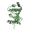
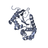
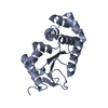
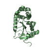
| Title | Structure and identification of a pterin dehydratase-like protein as a RuBisCO assembly factor in the alpha-carboxysome | ||||||
 Components Components | acRAF | ||||||
 Keywords Keywords | UNKNOWN FUNCTION / PCD / pterin-4a-carbinolamine dehydratase-like | ||||||
| Function / homology |  Function and homology information Function and homology information4a-hydroxytetrahydrobiopterin dehydratase / 4-alpha-hydroxytetrahydrobiopterin dehydratase activity / tetrahydrobiopterin biosynthetic process Similarity search - Function | ||||||
| Biological species |  Thiomonas intermedia (bacteria) Thiomonas intermedia (bacteria) | ||||||
| Method |  X-RAY DIFFRACTION / SYNCHROTRON / SAD / Resolution: 1.3 Å X-RAY DIFFRACTION / SYNCHROTRON / SAD / Resolution: 1.3 Å | ||||||
 Authors Authors | Wheatley, N.M. / Gidaniyan, S.D. / Cascio, D. / Yeates, T.O. | ||||||
 Citation Citation | Journal: J.Biol.Chem. / Year: 2014 Title: Structure and Identification of a Pterin Dehydratase-like Protein as a Ribulose-bisphosphate Carboxylase/Oxygenase (RuBisCO) Assembly Factor in the alpha-Carboxysome. Authors: Wheatley, N.M. / Sundberg, C.D. / Gidaniyan, S.D. / Cascio, D. / Yeates, T.O. | ||||||
| History |
|
- Structure visualization
| Structure viewer | Molecule: MolmilJmol/JSmol |
|---|
- Downloads & links
-Download
| PDBx/mmCIF format | 4low.cif.gz | 80.6 KB | Display | PDBx/mmCIF format |
|---|---|---|---|---|
| PDB format | pdb4low.ent.gz | 61.8 KB | Display | PDB format |
| PDBx/mmJSON format | 4low.json.gz | Tree view | PDBx/mmJSON format | |
| Others |  Other downloads Other downloads |
-Validation report
| Summary document | 4low_validation.pdf.gz | 454.6 KB | Display | wwPDB validaton report |
|---|---|---|---|---|
| Full document | 4low_full_validation.pdf.gz | 456.1 KB | Display | |
| Data in XML | 4low_validation.xml.gz | 10.1 KB | Display | |
| Data in CIF | 4low_validation.cif.gz | 13.4 KB | Display | |
| Arichive directory | https://data.pdbj.org/pub/pdb/validation_reports/lo/4lowftp://data.pdbj.org/pub/pdb/validation_reports/lo/4low | HTTPS FTP |
-Related structure data
| Similar structure data |
|---|


-Links
 PDBj
PDBj
- Assembly
| Deposited unit | 
| ||||||||
|---|---|---|---|---|---|---|---|---|---|
| 1 |
| ||||||||
| Unit cell |
|
-Components
| #1: Protein | Mass: 9720.723 Da / Num. of mol.: 2 / Fragment: UNP residues 7-88 Source method: isolated from a genetically manipulated source Source: (gene. exp.) Thiomonas intermedia (bacteria) / Strain: K12 / Gene: Tint_0125 / Plasmid: pET22b(+) / Production host: #2: Chemical | ChemComp-FMT /   Mass: 46.025 Da / Num. of mol.: 6 / Source method: obtained synthetically / Formula: CH2O2 Mass: 46.025 Da / Num. of mol.: 6 / Source method: obtained synthetically / Formula: CH2O2#3: Chemical |   Mass: 58.693 Da / Num. of mol.: 2 / Source method: obtained synthetically / Formula: Ni Mass: 58.693 Da / Num. of mol.: 2 / Source method: obtained synthetically / Formula: Ni#4: Water | ChemComp-HOH / |  Mass: 18.015 Da / Num. of mol.: 117 / Source method: isolated from a natural source / Formula: H2O Mass: 18.015 Da / Num. of mol.: 117 / Source method: isolated from a natural source / Formula: H2O |
|---|
-Experimental details
-Experiment
| Experiment | Method: X-RAY DIFFRACTION / Number of used crystals: 1 |
|---|
- Sample preparation
| Crystal | Density Matthews: 2.05 Å3/Da / Density % sol: 39.85 % |
|---|---|
| Crystal grow | Temperature: 298 K / Method: vapor diffusion, hanging drop Details: 17.5% PEG3350, 800 mM ammonium formate, VAPOR DIFFUSION, HANGING DROP, temperature 298K |
-Data collection
| Diffraction | Mean temperature: 100 K |
|---|---|
| Diffraction source | Source: SYNCHROTRON / Site: APS  / Beamline: 24-ID-C / Wavelength: 0.9791 Å / Beamline: 24-ID-C / Wavelength: 0.9791 Å |
| Detector | Type: DECTRIS PILATUS 6M / Detector: PIXEL / Date: Oct 27, 2012 |
| Radiation | Monochromator: Cryo-Cooled double crystal Si(111) / Protocol: SINGLE WAVELENGTH / Monochromatic (M) / Laue (L): M / Scattering type: x-ray |
| Radiation wavelength | Wavelength: 0.9791 Å / Relative weight: 1 |
| Reflection | Resolution: 1.3→51.11 Å / Num. obs: 36178 / % possible obs: 96.9 % |
- Processing
| Software |
| ||||||||||||||||||||||||||||||||||||||||||||||||||||||||||||||||||||||||||||||||||||||||||||||||||||||||||||
|---|---|---|---|---|---|---|---|---|---|---|---|---|---|---|---|---|---|---|---|---|---|---|---|---|---|---|---|---|---|---|---|---|---|---|---|---|---|---|---|---|---|---|---|---|---|---|---|---|---|---|---|---|---|---|---|---|---|---|---|---|---|---|---|---|---|---|---|---|---|---|---|---|---|---|---|---|---|---|---|---|---|---|---|---|---|---|---|---|---|---|---|---|---|---|---|---|---|---|---|---|---|---|---|---|---|---|---|---|---|
| Refinement | Method to determine structure: SAD / Resolution: 1.3→51.11 Å / Cor.coef. Fo:Fc: 0.951 / Cor.coef. Fo:Fc free: 0.9578 / Occupancy max: 1 / Occupancy min: 0.5 / SU R Cruickshank DPI: 0.059 / Cross valid method: THROUGHOUT / σ(F): 0 / SU R Blow DPI: 0.059 / SU Rfree Blow DPI: 0.054 / SU Rfree Cruickshank DPI: 0.054
| ||||||||||||||||||||||||||||||||||||||||||||||||||||||||||||||||||||||||||||||||||||||||||||||||||||||||||||
| Displacement parameters | Biso max: 174 Å2 / Biso mean: 32.3605 Å2 / Biso min: 13.88 Å2
| ||||||||||||||||||||||||||||||||||||||||||||||||||||||||||||||||||||||||||||||||||||||||||||||||||||||||||||
| Refine analyze | Luzzati coordinate error obs: 0.182 Å | ||||||||||||||||||||||||||||||||||||||||||||||||||||||||||||||||||||||||||||||||||||||||||||||||||||||||||||
| Refinement step | Cycle: LAST / Resolution: 1.3→51.11 Å
| ||||||||||||||||||||||||||||||||||||||||||||||||||||||||||||||||||||||||||||||||||||||||||||||||||||||||||||
| Refine LS restraints |
| ||||||||||||||||||||||||||||||||||||||||||||||||||||||||||||||||||||||||||||||||||||||||||||||||||||||||||||
| LS refinement shell | Resolution: 1.3→1.34 Å / Total num. of bins used: 18
| ||||||||||||||||||||||||||||||||||||||||||||||||||||||||||||||||||||||||||||||||||||||||||||||||||||||||||||
| Refinement TLS params. | Method: refined / Refine-ID: X-RAY DIFFRACTION
| ||||||||||||||||||||||||||||||||||||||||||||||||||||||||||||||||||||||||||||||||||||||||||||||||||||||||||||
| Refinement TLS group |
|
