 Movie
Movie Controller

[English] 日本語
 Yorodumi
- PDB-4lls: Crystal structure of a farnesyl diphosphate synthase from Roseoba... -
+ Open data

- Basic information
| Entry | Database: PDB / ID: 4lls | ||||||
|---|---|---|---|---|---|---|---|
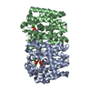
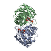
| Title | Crystal structure of a farnesyl diphosphate synthase from Roseobacter denitrificans OCh 114, target EFI-509393, with IPP, GSPP, and calcium bound in active site | ||||||
 Components Components | Geranyltranstransferase | ||||||
 Keywords Keywords | TRANSFERASE / Structural Genomics / Enzyme Function Initiative / polyprenyl synthetase | ||||||
| Function / homology |  Function and homology information Function and homology informationchlorophyll biosynthetic process / geranylgeranyl diphosphate synthase / carotenoid biosynthetic process / geranylgeranyl diphosphate synthase activity / photosynthesis / metal ion binding / cytoplasm Similarity search - Function | ||||||
| Biological species |  Roseobacter denitrificans (bacteria) Roseobacter denitrificans (bacteria) | ||||||
| Method |  X-RAY DIFFRACTION / SYNCHROTRON / MOLECULAR REPLACEMENT / Resolution: 1.5 Å X-RAY DIFFRACTION / SYNCHROTRON / MOLECULAR REPLACEMENT / Resolution: 1.5 Å | ||||||
 Authors Authors | Kim, J. / Toro, R. / Bhosle, R. / Al Obaidi, N.F. / Morisco, L.L. / Wasserman, S.R. / Sojitra, S. / Washington, E. / Scott Glenn, A. / Chowdhury, S. ...Kim, J. / Toro, R. / Bhosle, R. / Al Obaidi, N.F. / Morisco, L.L. / Wasserman, S.R. / Sojitra, S. / Washington, E. / Scott Glenn, A. / Chowdhury, S. / Evans, B. / Hammonds, J. / Hillerich, B. / Love, J. / Seidel, R.D. / Imker, H.J. / Stead, M. / Gerlt, J.A. / Almo, S.C. / Enzyme Function Initiative (EFI) | ||||||
 Citation Citation | Journal: To be Published Title: Crystal structure of a farnesyl diphosphate synthase from Roseobacter denitrificans OCh 114, target EFI-509393, with IPP, GSPP and calcium bound in active site Authors: Kim, J. / Toro, R. / Bhosle, R. / Al Obaidi, N.F. / Morisco, L.L. / Wasserman, S.R. / Sojitra, S. / Washington, E. / Scott Glenn, A. / Chowdhury, S. / Evans, B. / Hammonds, J. / Hillerich, B. ...Authors: Kim, J. / Toro, R. / Bhosle, R. / Al Obaidi, N.F. / Morisco, L.L. / Wasserman, S.R. / Sojitra, S. / Washington, E. / Scott Glenn, A. / Chowdhury, S. / Evans, B. / Hammonds, J. / Hillerich, B. / Love, J. / Seidel, R.D. / Imker, H.J. / Stead, M. / Gerlt, J.A. / Almo, S.C. | ||||||
| History |
|
- Structure visualization
| Structure viewer | Molecule: MolmilJmol/JSmol |
|---|
- Downloads & links
-Download
| PDBx/mmCIF format | 4lls.cif.gz | 137.2 KB | Display | PDBx/mmCIF format |
|---|---|---|---|---|
| PDB format | pdb4lls.ent.gz | 104.1 KB | Display | PDB format |
| PDBx/mmJSON format | 4lls.json.gz | Tree view | PDBx/mmJSON format | |
| Others |  Other downloads Other downloads |
-Validation report
| Arichive directory | https://data.pdbj.org/pub/pdb/validation_reports/ll/4llsftp://data.pdbj.org/pub/pdb/validation_reports/ll/4lls | HTTPS FTP |
|---|
-Related structure data
| Related structure data | 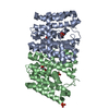 3lvsS S: Starting model for refinement |
|---|---|
| Similar structure data | |
| Other databases |


-Links
 PDBj
PDBj
- Assembly
| Deposited unit | 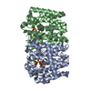
| ||||||||||||||||||
|---|---|---|---|---|---|---|---|---|---|---|---|---|---|---|---|---|---|---|---|
| 1 |
| ||||||||||||||||||
| Unit cell |
| ||||||||||||||||||
| Noncrystallographic symmetry (NCS) | NCS domain:
NCS domain segments: Component-ID: _ / Ens-ID: 1 / Beg auth comp-ID: SER / Beg label comp-ID: SER / End auth comp-ID: THR / End label comp-ID: THR / Refine code: _ / Auth seq-ID: 0 - 288 / Label seq-ID: 22 - 310
|
-Components
| #1: Protein | Mass: 32847.074 Da / Num. of mol.: 2 Source method: isolated from a genetically manipulated source Source: (gene. exp.) Roseobacter denitrificans (bacteria) / Strain: ATCC 33942 / OCh 114 / Gene: ispA, RD1_0549 / Production host: References: UniProt: Q16CN9, (2E,6E)-farnesyl diphosphate synthase #2: Chemical |   Mass: 246.092 Da / Num. of mol.: 2 / Source method: obtained synthetically / Formula: C5H12O7P2 Mass: 246.092 Da / Num. of mol.: 2 / Source method: obtained synthetically / Formula: C5H12O7P2#3: Chemical |   Mass: 330.275 Da / Num. of mol.: 2 / Source method: obtained synthetically / Formula: C10H20O6P2S Mass: 330.275 Da / Num. of mol.: 2 / Source method: obtained synthetically / Formula: C10H20O6P2S#4: Chemical | ChemComp-CA /   Mass: 40.078 Da / Num. of mol.: 6 / Source method: obtained synthetically / Formula: Ca Mass: 40.078 Da / Num. of mol.: 6 / Source method: obtained synthetically / Formula: Ca#5: Water | ChemComp-HOH / |  Mass: 18.015 Da / Num. of mol.: 435 / Source method: isolated from a natural source / Formula: H2O Mass: 18.015 Da / Num. of mol.: 435 / Source method: isolated from a natural source / Formula: H2O |
|---|
-Experimental details
-Experiment
| Experiment | Method: X-RAY DIFFRACTION / Number of used crystals: 1 |
|---|
- Sample preparation
| Crystal | Density Matthews: 2.18 Å3/Da / Density % sol: 43.61 % |
|---|---|
| Crystal grow | Temperature: 293 K / Method: vapor diffusion, sitting drop / pH: 6 Details: 0.2 M calcium acetate, 0.1 M MES/NaOH, pH 6.0, 20% v/v PEG8000, VAPOR DIFFUSION, SITTING DROP, temperature 293K |
-Data collection
| Diffraction | Mean temperature: 100 K |
|---|---|
| Diffraction source | Source: SYNCHROTRON / Site: APS  / Beamline: 31-ID / Wavelength: 0.979 Å / Beamline: 31-ID / Wavelength: 0.979 Å |
| Detector | Type: RAYONIX MX225HE / Detector: CCD / Date: Jun 14, 2013 |
| Radiation | Monochromator: diamond(111) / Protocol: SINGLE WAVELENGTH / Monochromatic (M) / Laue (L): M / Scattering type: x-ray |
| Radiation wavelength | Wavelength: 0.979 Å / Relative weight: 1 |
| Reflection | Resolution: 1.5→50 Å / Num. all: 90012 / Num. obs: 89715 / % possible obs: 99.7 % / Redundancy: 3.8 % / Rmerge(I) obs: 0.074 / Net I/σ(I): 15.2 |
| Reflection shell | Resolution: 1.5→1.53 Å / Redundancy: 3.7 % / Rmerge(I) obs: 0.632 / Mean I/σ(I) obs: 2.1 / % possible all: 99.9 |
- Processing
| Software |
| ||||||||||||||||||||||||||||||||||||||||||||||||||||||||||||||||||||||||||||||||||||||||||||||||||||||||||||||||||||||||||||||||||||||||||||||||||||||||||||||||||||||||||||||||||||||
|---|---|---|---|---|---|---|---|---|---|---|---|---|---|---|---|---|---|---|---|---|---|---|---|---|---|---|---|---|---|---|---|---|---|---|---|---|---|---|---|---|---|---|---|---|---|---|---|---|---|---|---|---|---|---|---|---|---|---|---|---|---|---|---|---|---|---|---|---|---|---|---|---|---|---|---|---|---|---|---|---|---|---|---|---|---|---|---|---|---|---|---|---|---|---|---|---|---|---|---|---|---|---|---|---|---|---|---|---|---|---|---|---|---|---|---|---|---|---|---|---|---|---|---|---|---|---|---|---|---|---|---|---|---|---|---|---|---|---|---|---|---|---|---|---|---|---|---|---|---|---|---|---|---|---|---|---|---|---|---|---|---|---|---|---|---|---|---|---|---|---|---|---|---|---|---|---|---|---|---|---|---|---|---|
| Refinement | Method to determine structure: MOLECULAR REPLACEMENT Starting model: PDB ENTRY 3LVS Resolution: 1.5→26.5 Å / Cor.coef. Fo:Fc: 0.968 / Cor.coef. Fo:Fc free: 0.96 / SU B: 1.236 / SU ML: 0.047 / Cross valid method: THROUGHOUT / ESU R: 0.07 / ESU R Free: 0.072 / Stereochemistry target values: MAXIMUM LIKELIHOOD Details: HYDROGENS HAVE BEEN ADDED IN THE RIDING POSITIONS U VALUES : REFINED INDIVIDUALLY
| ||||||||||||||||||||||||||||||||||||||||||||||||||||||||||||||||||||||||||||||||||||||||||||||||||||||||||||||||||||||||||||||||||||||||||||||||||||||||||||||||||||||||||||||||||||||
| Solvent computation | Ion probe radii: 0.8 Å / Shrinkage radii: 0.8 Å / VDW probe radii: 1.2 Å / Solvent model: MASK | ||||||||||||||||||||||||||||||||||||||||||||||||||||||||||||||||||||||||||||||||||||||||||||||||||||||||||||||||||||||||||||||||||||||||||||||||||||||||||||||||||||||||||||||||||||||
| Displacement parameters | Biso mean: 21.757 Å2
| ||||||||||||||||||||||||||||||||||||||||||||||||||||||||||||||||||||||||||||||||||||||||||||||||||||||||||||||||||||||||||||||||||||||||||||||||||||||||||||||||||||||||||||||||||||||
| Refinement step | Cycle: LAST / Resolution: 1.5→26.5 Å
| ||||||||||||||||||||||||||||||||||||||||||||||||||||||||||||||||||||||||||||||||||||||||||||||||||||||||||||||||||||||||||||||||||||||||||||||||||||||||||||||||||||||||||||||||||||||
| Refine LS restraints |
|