| Entry | Database: PDB / ID: 4lh7
|
|---|
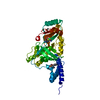
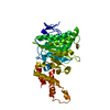
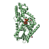
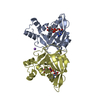
| Title | Crystal structure of a LigA inhibitor |
|---|
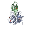
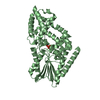
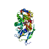
 Components Components | DNA ligase |
|---|
 Keywords Keywords | LIGASE/LIGASE INHIBITOR / Protein-inhibitor complex / LIGASE-LIGASE INHIBITOR complex |
|---|
| Function / homology |  Function and homology information Function and homology information
Dna Ligase; domain 1 - #70 / Zinc-finger, NAD-dependent DNA ligase C4-type / NAD-dependent DNA ligase C4 zinc finger domain / NAD-dependent DNA ligase, active site / NAD-dependent DNA ligase, conserved site / NAD-dependent DNA ligase signature 1. / NAD-dependent DNA ligase signature 2. / NAD-dependent DNA ligase / NAD-dependent DNA ligase, OB-fold / NAD-dependent DNA ligase, adenylation ...Dna Ligase; domain 1 - #70 / Zinc-finger, NAD-dependent DNA ligase C4-type / NAD-dependent DNA ligase C4 zinc finger domain / NAD-dependent DNA ligase, active site / NAD-dependent DNA ligase, conserved site / NAD-dependent DNA ligase signature 1. / NAD-dependent DNA ligase signature 2. / NAD-dependent DNA ligase / NAD-dependent DNA ligase, OB-fold / NAD-dependent DNA ligase, adenylation / NAD-dependent DNA ligase, N-terminal / NAD-dependent DNA ligase adenylation domain / NAD-dependent DNA ligase OB-fold domain / Ligase N family / DisA/LigA, helix-hairpin-helix motif / Helix-hairpin-helix motif / DNA ligase/mRNA capping enzyme / Helix hairpin bin / RuvA domain 2-like / BRCA1 C Terminus (BRCT) domain / Helix-hairpin-helix domain / breast cancer carboxy-terminal domain / D-amino Acid Aminotransferase; Chain A, domain 1 / BRCT domain profile. / BRCT domain / BRCT domain superfamily / Dna Ligase; domain 1 / Helix Hairpins / Nucleic acid-binding, OB-fold / 2-Layer Sandwich / Orthogonal Bundle / Mainly Alpha / Alpha BetaSimilarity search - Domain/homology |
|---|
| Biological species |   Enterococcus faecalis (bacteria) Enterococcus faecalis (bacteria) |
|---|
| Method |  X-RAY DIFFRACTION / MOLECULAR REPLACEMENT / Resolution: 1.9 Å X-RAY DIFFRACTION / MOLECULAR REPLACEMENT / Resolution: 1.9 Å |
|---|
 Authors Authors | Boriack-Sjodin, P.A. / Prince, D.B. |
|---|
 Citation Citation | Journal: Bioorg.Med.Chem.Lett. / Year: 2014
Title: Identification through structure-based methods of a bacterial NAD(+)-dependent DNA ligase inhibitor that avoids known resistance mutations.
Authors: Murphy-Benenato, K. / Wang, H. / McGuire, H.M. / Davis, H.E. / Gao, N. / Prince, D.B. / Jahic, H. / Stokes, S.S. / Boriack-Sjodin, P.A. |
|---|
| History | | Deposition | Jun 30, 2013 | Deposition site: RCSB / Processing site: RCSB |
|---|
| Revision 1.0 | Dec 25, 2013 | Provider: repository / Type: Initial release |
|---|
| Revision 1.1 | Jan 8, 2014 | Group: Database references |
|---|
| Revision 1.2 | Jan 13, 2021 | Group: Derived calculations / Source and taxonomy
Category: entity_src_gen / pdbx_struct_conn_angle ...entity_src_gen / pdbx_struct_conn_angle / struct_conn / struct_site
Item: _entity_src_gen.pdbx_host_org_ncbi_taxonomy_id / _pdbx_struct_conn_angle.ptnr1_auth_comp_id ..._entity_src_gen.pdbx_host_org_ncbi_taxonomy_id / _pdbx_struct_conn_angle.ptnr1_auth_comp_id / _pdbx_struct_conn_angle.ptnr1_auth_seq_id / _pdbx_struct_conn_angle.ptnr1_label_asym_id / _pdbx_struct_conn_angle.ptnr1_label_comp_id / _pdbx_struct_conn_angle.ptnr1_label_seq_id / _pdbx_struct_conn_angle.ptnr3_auth_comp_id / _pdbx_struct_conn_angle.ptnr3_auth_seq_id / _pdbx_struct_conn_angle.ptnr3_label_asym_id / _pdbx_struct_conn_angle.ptnr3_label_comp_id / _pdbx_struct_conn_angle.ptnr3_label_seq_id / _pdbx_struct_conn_angle.value / _struct_conn.pdbx_dist_value / _struct_conn.ptnr1_auth_comp_id / _struct_conn.ptnr1_auth_seq_id / _struct_conn.ptnr1_label_asym_id / _struct_conn.ptnr1_label_atom_id / _struct_conn.ptnr1_label_comp_id / _struct_conn.ptnr1_label_seq_id / _struct_conn.ptnr2_auth_comp_id / _struct_conn.ptnr2_auth_seq_id / _struct_conn.ptnr2_label_asym_id / _struct_conn.ptnr2_label_atom_id / _struct_conn.ptnr2_label_comp_id / _struct_site.pdbx_auth_asym_id / _struct_site.pdbx_auth_comp_id / _struct_site.pdbx_auth_seq_id |
|---|
| Revision 1.3 | Feb 28, 2024 | Group: Data collection / Database references / Category: chem_comp_atom / chem_comp_bond / database_2
Item: _database_2.pdbx_DOI / _database_2.pdbx_database_accession |
|---|
|
|---|
 Movie
Movie Controller


 Open data
 Basic information
 Structure visualization
 Downloads & links
 Other downloads


 PDBj
PDBj


 Assembly
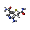
 Mass: 236.250 Da / Num. of mol.: 1 / Source method: obtained synthetically / Formula: C9H8N4O2S
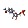
 Mass: 335.227 Da / Num. of mol.: 1 / Source method: obtained synthetically / Formula: C11H16N2O8P
 Mass: 22.990 Da / Num. of mol.: 2 / Source method: obtained synthetically / Formula: Na
 Mass: 18.015 Da / Num. of mol.: 159 / Source method: isolated from a natural source / Formula: H2O
 Sample preparation
 Processing