 Movie
Movie Controller

[English] 日本語
 Yorodumi
- PDB-4lfm: Crystal Structure of D-galactose-6-phosphate isomerase in complex... -
+ Open data

- Basic information
| Entry | Database: PDB / ID: 4lfm | ||||||
|---|---|---|---|---|---|---|---|
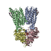
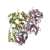
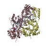
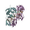
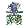
| Title | Crystal Structure of D-galactose-6-phosphate isomerase in complex with D-psicose | ||||||
 Components Components |
| ||||||
 Keywords Keywords | ISOMERASE / Rossmann-like alpha-beta-alpha sandwich fold / Rossmann Fold / Sugar-phosphate Binding / Isomerization | ||||||
| Function / homology | Ribose 5-phosphate Isomerase B; Chain: A, / Sugar-phosphate isomerase, RpiB/LacA/LacB / 3-Layer(aba) Sandwich / Alpha Beta / D-psicose / : / :  Function and homology information Function and homology information | ||||||
| Biological species |  Lactobacillus rhamnosus (bacteria) Lactobacillus rhamnosus (bacteria) | ||||||
| Method |  X-RAY DIFFRACTION / SYNCHROTRON / MOLECULAR REPLACEMENT / Resolution: 1.65 Å X-RAY DIFFRACTION / SYNCHROTRON / MOLECULAR REPLACEMENT / Resolution: 1.65 Å | ||||||
 Authors Authors | Jung, W.S. / Pan, C.H. | ||||||
 Citation Citation | Journal: Plos One / Year: 2013 Title: Crystal structure and substrate specificity of D-galactose-6-phosphate isomerase complexed with substrates. Authors: Jung, W.S. / Singh, R.K. / Lee, J.K. / Pan, C.H. | ||||||
| History |
|
- Structure visualization
| Structure viewer | Molecule: MolmilJmol/JSmol |
|---|
- Downloads & links
-Download
| PDBx/mmCIF format | 4lfm.cif.gz | 134.5 KB | Display | PDBx/mmCIF format |
|---|---|---|---|---|
| PDB format | pdb4lfm.ent.gz | 104.6 KB | Display | PDB format |
| PDBx/mmJSON format | 4lfm.json.gz | Tree view | PDBx/mmJSON format | |
| Others |  Other downloads Other downloads |
-Validation report
| Arichive directory | https://data.pdbj.org/pub/pdb/validation_reports/lf/4lfmftp://data.pdbj.org/pub/pdb/validation_reports/lf/4lfm | HTTPS FTP |
|---|
-Related structure data
| Related structure data |  4lfkSC 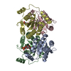 4lflC  4lfnC S: Starting model for refinement C: citing same article ( |
|---|---|
| Similar structure data |


-Links
 PDBj
PDBj- Assembly
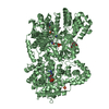
| Deposited unit | 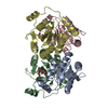
| ||||||||
|---|---|---|---|---|---|---|---|---|---|
| 1 | 
| ||||||||
| Unit cell |
|
-Components
| #1: Protein | Mass: 17884.320 Da / Num. of mol.: 2 Source method: isolated from a genetically manipulated source Source: (gene. exp.) Lactobacillus rhamnosus (bacteria) / Strain: Lc 705 / Gene: lacA, LC705_00641 / Plasmid: pET28 / Production host: #2: Protein | Mass: 18889.338 Da / Num. of mol.: 2 Source method: isolated from a genetically manipulated source Source: (gene. exp.) Lactobacillus rhamnosus (bacteria) / Strain: Lc 705 / Gene: lacB, LC705_00640 / Plasmid: pET28 / Production host: #3: Sugar | 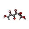  Type: D-saccharide / Mass: 180.156 Da / Num. of mol.: 2 Type: D-saccharide / Mass: 180.156 Da / Num. of mol.: 2Source method: isolated from a genetically manipulated source Formula: C6H12O6 #4: Water | ChemComp-HOH / |  Mass: 18.015 Da / Num. of mol.: 199 / Source method: isolated from a natural source / Formula: H2O Mass: 18.015 Da / Num. of mol.: 199 / Source method: isolated from a natural source / Formula: H2O |
|---|
-Experimental details
-Experiment
| Experiment | Method: X-RAY DIFFRACTION / Number of used crystals: 1 |
|---|
- Sample preparation
| Crystal | Density Matthews: 2.34 Å3/Da / Density % sol: 47.33 % / Mosaicity: 0.607 ° |
|---|---|
| Crystal grow | Temperature: 295 K / Method: vapor diffusion, hanging drop Details: 20% PEG 3350, 0.2M potassium formate, VAPOR DIFFUSION, HANGING DROP, temperature 295K |
-Data collection
| Diffraction | Mean temperature: 100 K | |||||||||||||||||||||||||||||||||||||||||||||||||||||||||||||||||||||||||||||||||||||||||||||||||||||||||||||||||||||||||||||||||||||||||||||||||||
|---|---|---|---|---|---|---|---|---|---|---|---|---|---|---|---|---|---|---|---|---|---|---|---|---|---|---|---|---|---|---|---|---|---|---|---|---|---|---|---|---|---|---|---|---|---|---|---|---|---|---|---|---|---|---|---|---|---|---|---|---|---|---|---|---|---|---|---|---|---|---|---|---|---|---|---|---|---|---|---|---|---|---|---|---|---|---|---|---|---|---|---|---|---|---|---|---|---|---|---|---|---|---|---|---|---|---|---|---|---|---|---|---|---|---|---|---|---|---|---|---|---|---|---|---|---|---|---|---|---|---|---|---|---|---|---|---|---|---|---|---|---|---|---|---|---|---|---|---|
| Diffraction source | Source: SYNCHROTRON / Site: PAL/PLS  / Beamline: 7A (6B, 6C1) / Wavelength: 0.97932 Å / Beamline: 7A (6B, 6C1) / Wavelength: 0.97932 Å | |||||||||||||||||||||||||||||||||||||||||||||||||||||||||||||||||||||||||||||||||||||||||||||||||||||||||||||||||||||||||||||||||||||||||||||||||||
| Detector | Type: ADSC QUANTUM 270 / Detector: CCD / Date: Jun 26, 2012 | |||||||||||||||||||||||||||||||||||||||||||||||||||||||||||||||||||||||||||||||||||||||||||||||||||||||||||||||||||||||||||||||||||||||||||||||||||
| Radiation | Protocol: SINGLE WAVELENGTH / Monochromatic (M) / Laue (L): M / Scattering type: x-ray | |||||||||||||||||||||||||||||||||||||||||||||||||||||||||||||||||||||||||||||||||||||||||||||||||||||||||||||||||||||||||||||||||||||||||||||||||||
| Radiation wavelength | Wavelength: 0.97932 Å / Relative weight: 1 | |||||||||||||||||||||||||||||||||||||||||||||||||||||||||||||||||||||||||||||||||||||||||||||||||||||||||||||||||||||||||||||||||||||||||||||||||||
| Reflection | Resolution: 1.65→50 Å / Num. obs: 82373 / % possible obs: 98.5 % / Redundancy: 12.9 % / Rmerge(I) obs: 0.096 / Χ2: 1.268 / Net I/σ(I): 8.9 | |||||||||||||||||||||||||||||||||||||||||||||||||||||||||||||||||||||||||||||||||||||||||||||||||||||||||||||||||||||||||||||||||||||||||||||||||||
| Reflection shell |
|
- Processing
| Software |
| ||||||||||||||||||||||||||||
|---|---|---|---|---|---|---|---|---|---|---|---|---|---|---|---|---|---|---|---|---|---|---|---|---|---|---|---|---|---|
| Refinement | Method to determine structure: MOLECULAR REPLACEMENT Starting model: 4LFK Resolution: 1.65→50 Å / Occupancy max: 1 / Occupancy min: 1 / σ(F): 2
| ||||||||||||||||||||||||||||
| Solvent computation | Bsol: 23.67 Å2 | ||||||||||||||||||||||||||||
| Displacement parameters | Biso max: 58.59 Å2 / Biso mean: 24.0015 Å2 / Biso min: 11.38 Å2
| ||||||||||||||||||||||||||||
| Refinement step | Cycle: LAST / Resolution: 1.65→50 Å
| ||||||||||||||||||||||||||||
| Refine LS restraints |
| ||||||||||||||||||||||||||||
| Xplor file |
|
