 Movie
Movie Controller

[English] 日本語
 Yorodumi
- PDB-4lcz: Crystal structure of a multilayer-packed major light-harvesting c... -
+ Open data

- Basic information
| Entry | Database: PDB / ID: 4lcz | |||||||||
|---|---|---|---|---|---|---|---|---|---|---|
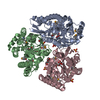
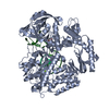
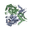
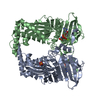
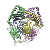
| Title | Crystal structure of a multilayer-packed major light-harvesting complex | |||||||||
 Components Components | Major chlorophyll a/b binding protein LHCb1.3 | |||||||||
 Keywords Keywords | MEMBRANE PROTEIN / Light collecting / Photon protection / Grana stacking / Chlorophyll binding | |||||||||
| Function / homology |  Function and homology information Function and homology informationphotosynthesis, light harvesting in photosystem I / photosynthesis, light harvesting / photosystem I / photosystem II / chlorophyll binding / chloroplast thylakoid membrane / response to light stimulus / metal ion binding Similarity search - Function | |||||||||
| Biological species |  Spinacia oleracea (spinach) Spinacia oleracea (spinach) | |||||||||
| Method |  X-RAY DIFFRACTION / SYNCHROTRON / MOLECULAR REPLACEMENT / Resolution: 2.6 Å X-RAY DIFFRACTION / SYNCHROTRON / MOLECULAR REPLACEMENT / Resolution: 2.6 Å | |||||||||
 Authors Authors | Wan, T. / Li, M. / Chang, W.R. | |||||||||
 Citation Citation | Journal: Mol Plant / Year: 2014 Title: Crystal structure of a multilayer packed major light-harvesting complex: implications for grana stacking in higher plants. Authors: Wan, T. / Li, M. / Zhao, X. / Zhang, J. / Liu, Z. / Chang, W. | |||||||||
| History |
|
- Structure visualization
| Structure viewer | Molecule: MolmilJmol/JSmol |
|---|
- Downloads & links
-Download
| PDBx/mmCIF format | 4lcz.cif.gz | 421 KB | Display | PDBx/mmCIF format |
|---|---|---|---|---|
| PDB format | pdb4lcz.ent.gz | 357.7 KB | Display | PDB format |
| PDBx/mmJSON format | 4lcz.json.gz | Tree view | PDBx/mmJSON format | |
| Others |  Other downloads Other downloads |
-Validation report
| Arichive directory | https://data.pdbj.org/pub/pdb/validation_reports/lc/4lczftp://data.pdbj.org/pub/pdb/validation_reports/lc/4lcz | HTTPS FTP |
|---|
-Related structure data
| Related structure data | 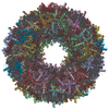 1rwtS S: Starting model for refinement |
|---|---|
| Similar structure data |


-Links
 PDBj
PDBj


- Assembly
| Deposited unit | 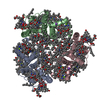
| ||||||||
|---|---|---|---|---|---|---|---|---|---|
| 1 |
| ||||||||
| Unit cell |
| ||||||||
| Components on special symmetry positions |
|
-Components
-Protein , 1 types, 3 molecules ABC
| #1: Protein | Mass: 24129.248 Da / Num. of mol.: 3 / Fragment: UNP residues 44-267 / Source method: isolated from a natural source / Source: (natural) Spinacia oleracea (spinach) / References: UniProt: B3WFZ6, UniProt: P12333*PLUS |
|---|
-Non-polymers , 9 types, 223 molecules 


| #2: Chemical | ChemComp-LUT / (  Mass: 568.871 Da / Num. of mol.: 6 / Source method: obtained synthetically / Formula: C40H56O2 Mass: 568.871 Da / Num. of mol.: 6 / Source method: obtained synthetically / Formula: C40H56O2#3: Chemical |  Mass: 600.870 Da / Num. of mol.: 3 / Source method: obtained synthetically / Formula: C40H56O4 Mass: 600.870 Da / Num. of mol.: 3 / Source method: obtained synthetically / Formula: C40H56O4#4: Chemical |  Mass: 722.970 Da / Num. of mol.: 3 / Source method: obtained synthetically / Formula: C38H75O10P / Comment: phospholipid*YM Mass: 722.970 Da / Num. of mol.: 3 / Source method: obtained synthetically / Formula: C38H75O10P / Comment: phospholipid*YM#5: Chemical | ChemComp-CHL /  Mass: 907.472 Da / Num. of mol.: 18 / Source method: obtained synthetically / Formula: C55H70MgN4O6 Mass: 907.472 Da / Num. of mol.: 18 / Source method: obtained synthetically / Formula: C55H70MgN4O6#6: Chemical | ChemComp-CLA /  Mass: 893.489 Da / Num. of mol.: 24 / Source method: obtained synthetically / Formula: C55H72MgN4O5 Mass: 893.489 Da / Num. of mol.: 24 / Source method: obtained synthetically / Formula: C55H72MgN4O5#7: Chemical |  Mass: 136.989 Da / Num. of mol.: 3 / Source method: obtained synthetically / Formula: C2H6AsO2 Mass: 136.989 Da / Num. of mol.: 3 / Source method: obtained synthetically / Formula: C2H6AsO2#8: Chemical | ChemComp-ZN /  Mass: 65.409 Da / Num. of mol.: 5 / Source method: obtained synthetically / Formula: Zn Mass: 65.409 Da / Num. of mol.: 5 / Source method: obtained synthetically / Formula: Zn#9: Chemical | ChemComp-NA /  Mass: 22.990 Da / Num. of mol.: 5 / Source method: obtained synthetically / Formula: Na Mass: 22.990 Da / Num. of mol.: 5 / Source method: obtained synthetically / Formula: Na#10: Water | ChemComp-HOH / | Mass: 18.015 Da / Num. of mol.: 156 / Source method: isolated from a natural source / Formula: H2O |
|---|
-Experimental details
-Experiment
| Experiment | Method: X-RAY DIFFRACTION / Number of used crystals: 1 |
|---|
- Sample preparation
| Crystal | Density Matthews: 7.97 Å3/Da / Density % sol: 84.58 % |
|---|---|
| Crystal grow | Temperature: 291 K / pH: 6.5 Details: 2 mg/ml DGDG, 100mM di-sodium cacodylate, 10-12%(w/v) PEG2000, 20-50mM zinc acetate , pH 6.5, VAPOR DIFFUSION, SITTING DROP, temperature 291K |
-Data collection
| Diffraction | Mean temperature: 173 K |
|---|---|
| Diffraction source | Source: SYNCHROTRON / Site: SSRF  / Beamline: BL17U / Wavelength: 0.96 / Beamline: BL17U / Wavelength: 0.96 |
| Detector | Type: ADSC QUANTUM 315r / Detector: CCD / Date: Dec 13, 2012 |
| Radiation | Monochromator: GRAPHITE / Protocol: SINGLE WAVELENGTH / Monochromatic (M) / Laue (L): M / Scattering type: x-ray |
| Radiation wavelength | Wavelength: 0.96 Å / Relative weight: 1 |
| Reflection | Resolution: 2.6→50 Å / Num. obs: 69844 / % possible obs: 100 % / Observed criterion σ(I): 1 / Redundancy: 3.7 % / Biso Wilson estimate: 30.6 Å2 / Rmerge(I) obs: 0.082 / Net I/σ(I): 15.2 |
| Reflection shell | Resolution: 2.6→2.69 Å / Redundancy: 3.6 % / Rmerge(I) obs: 0.761 / Mean I/σ(I) obs: 1.8 / Rsym value: 0.643 / % possible all: 98.1 |
- Processing
| Software |
| |||||||||||||||||||||||||||||||||||||||||||||||||||||||||||||||||||||||||||||||||||||||||||||||||||||||||||||||||||||||||||||||||||||||||||||||||||||||||||||||||
|---|---|---|---|---|---|---|---|---|---|---|---|---|---|---|---|---|---|---|---|---|---|---|---|---|---|---|---|---|---|---|---|---|---|---|---|---|---|---|---|---|---|---|---|---|---|---|---|---|---|---|---|---|---|---|---|---|---|---|---|---|---|---|---|---|---|---|---|---|---|---|---|---|---|---|---|---|---|---|---|---|---|---|---|---|---|---|---|---|---|---|---|---|---|---|---|---|---|---|---|---|---|---|---|---|---|---|---|---|---|---|---|---|---|---|---|---|---|---|---|---|---|---|---|---|---|---|---|---|---|---|---|---|---|---|---|---|---|---|---|---|---|---|---|---|---|---|---|---|---|---|---|---|---|---|---|---|---|---|---|---|---|---|
| Refinement | Method to determine structure: MOLECULAR REPLACEMENT Starting model: 1RWT Resolution: 2.6→43.45 Å / SU ML: 0.26 / σ(F): 1.35 / Phase error: 26.38 / Stereochemistry target values: ML Details: MATTHEW COEFFICIENT AND SOLVENT CONTENT HAVE BEEN CALCULATED BY SFCHECK CONTAINED IN THE CCP4 SUITE.
| |||||||||||||||||||||||||||||||||||||||||||||||||||||||||||||||||||||||||||||||||||||||||||||||||||||||||||||||||||||||||||||||||||||||||||||||||||||||||||||||||
| Solvent computation | Shrinkage radii: 0.86 Å / VDW probe radii: 1.1 Å / Solvent model: FLAT BULK SOLVENT MODEL / Bsol: 60 Å2 / ksol: 0.4 e/Å3 | |||||||||||||||||||||||||||||||||||||||||||||||||||||||||||||||||||||||||||||||||||||||||||||||||||||||||||||||||||||||||||||||||||||||||||||||||||||||||||||||||
| Displacement parameters |
| |||||||||||||||||||||||||||||||||||||||||||||||||||||||||||||||||||||||||||||||||||||||||||||||||||||||||||||||||||||||||||||||||||||||||||||||||||||||||||||||||
| Refinement step | Cycle: LAST / Resolution: 2.6→43.45 Å
| |||||||||||||||||||||||||||||||||||||||||||||||||||||||||||||||||||||||||||||||||||||||||||||||||||||||||||||||||||||||||||||||||||||||||||||||||||||||||||||||||
| Refine LS restraints |
| |||||||||||||||||||||||||||||||||||||||||||||||||||||||||||||||||||||||||||||||||||||||||||||||||||||||||||||||||||||||||||||||||||||||||||||||||||||||||||||||||
| LS refinement shell |
| |||||||||||||||||||||||||||||||||||||||||||||||||||||||||||||||||||||||||||||||||||||||||||||||||||||||||||||||||||||||||||||||||||||||||||||||||||||||||||||||||
| Refinement TLS params. | Method: refined / Origin x: 11.4897 Å / Origin y: 53.4186 Å / Origin z: 24.9062 Å
| |||||||||||||||||||||||||||||||||||||||||||||||||||||||||||||||||||||||||||||||||||||||||||||||||||||||||||||||||||||||||||||||||||||||||||||||||||||||||||||||||
| Refinement TLS group |
|
