- PDB-4lb8: Crystal structure of a DUF4848 family protein (BT3222) from Bacte... -
+
Open data
ID or keywords:
Loading...
-
Basic information
Entry
Database: PDB / ID: 4lb8
Title
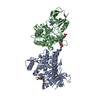
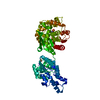
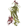
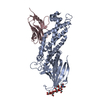
Crystal structure of a DUF4848 family protein (BT3222) from Bacteroides thetaiotaomicron VPI-5482 at 2.49 A resolution
Components
Uncharacterized protein
Keywords
STRUCTURAL GENOMICS / UNKNOWN FUNCTION / PF16140 family protein / DUF4848 / Joint Center for Structural Genomics / JCSG / Protein Structure Initiative / PSI-BIOLOGY
Function / homology
Immunoglobulin-like - #3900 / Protein of unknown function DUF4848 / Domain of unknown function (DUF4848) / Prokaryotic membrane lipoprotein lipid attachment site profile. / Immunoglobulin-like / Sandwich / Mainly Beta / DI(HYDROXYETHYL)ETHER / DUF4848 domain-containing protein
Function and homology information
Biological species
Bacteroides thetaiotaomicron (bacteria)
Method
X-RAY DIFFRACTION / SYNCHROTRON / MAD / Resolution: 2.49 Å
Mass: 18.015 Da / Num. of mol.: 79 / Source method: isolated from a natural source / Formula: H2O
Has protein modification
Y
Sequence details
THIS CONSTRUCT WAS EXPRESSED WITH A PURIFICATION TAG MGSDKIHHHHHHENLYFQG. THE TAG WAS REMOVED WITH ...THIS CONSTRUCT WAS EXPRESSED WITH A PURIFICATION TAG MGSDKIHHHHHHENLYFQG. THE TAG WAS REMOVED WITH TEV PROTEASE LEAVING ONLY A GLYCINE (0) FOLLOWED BY RESIDUES 22-310 OF THE TARGET SEQUENCE.
-
Experimental details
-
Experiment
Experiment
Method: X-RAY DIFFRACTION / Number of used crystals: 1
-
Sample preparation
Crystal
Density Matthews: 3.06 Å3/Da / Density % sol: 59.8 %
Type: DECTRIS PILATUS 6M / Detector: PIXEL / Date: May 2, 2013 Details: Flat mirror (vertical focusing); single crystal Si(111) bent monochromator (horizontal focusing)
Radiation
Monochromator: single crystal Si(111) bent / Protocol: MAD / Monochromatic (M) / Laue (L): M / Scattering type: x-ray
Radiation wavelength
ID
Wavelength (Å)
Relative weight
1
0.91162
1
2
0.9786
1
3
0.97805
1
Reflection
Resolution: 2.49→29.357 Å / Num. obs: 14384 / % possible obs: 95.2 % / Observed criterion σ(I): -3 / Biso Wilson estimate: 59.579 Å2 / Rmerge(I) obs: 0.067 / Net I/σ(I): 8.96
Reflection shell
Resolution (Å)
Rmerge(I) obs
Mean I/σ(I) obs
Num. measured obs
Num. unique obs
Diffraction-ID
% possible all
2.49-2.58
0.634
1.2
4653
2622
1
94
2.58-2.68
0.423
1.7
4208
2501
1
93.4
2.68-2.8
0.313
2.4
4904
2647
1
97.5
2.8-2.95
0.235
3.2
5063
2741
1
97
2.95-3.14
0.147
4.8
5072
2744
1
96.2
3.14-3.38
0.097
7.4
4634
2589
1
94.2
3.38-3.71
0.058
11
4567
2542
1
94.7
3.71-4.25
0.04
15.6
5033
2730
1
97
4.25-5.33
0.031
19.8
4536
2550
1
93.3
5.33-29.357
0.029
22.5
4972
2686
1
94.4
-
Phasing
Phasing
Method: MAD
-
Processing
Software
Name
Version
Classification
NB
MolProbity
3beta29
modelbuilding
PDB_EXTRACT
3.1
dataextraction
SOLVE
phasing
XSCALE
July4, 2012
datascaling
BUSTER-TNT
refinement
XDS
datareduction
BUSTER
2.10.0
refinement
Refinement
Method to determine structure: MAD / Resolution: 2.49→29.357 Å / Cor.coef. Fo:Fc: 0.9357 / Cor.coef. Fo:Fc free: 0.8971 / Occupancy max: 1 / Occupancy min: 0.5 / Cross valid method: THROUGHOUT / σ(F): 0 Details: 1.A MET-INHIBITION PROTOCOL WAS USED FOR SELENOMETHIONINE INCORPORATION DURING PROTEIN EXPRESSION. THE OCCUPANCY OF THE SE ATOMS IN THE MSE RESIDUES WAS REDUCED TO 0.75 FOR THE REDUCED ...Details: 1.A MET-INHIBITION PROTOCOL WAS USED FOR SELENOMETHIONINE INCORPORATION DURING PROTEIN EXPRESSION. THE OCCUPANCY OF THE SE ATOMS IN THE MSE RESIDUES WAS REDUCED TO 0.75 FOR THE REDUCED SCATTERING POWER DUE TO PARTIAL S-MET INCORPORATION. 2.ATOM RECORD CONTAINS SUM OF TLS AND RESIDUAL B FACTORS. 3.ANISOU RECORD CONTAINS SUM OF TLS AND RESIDUAL U FACTORS. 4.CHLORIDE (CL) AND PEG3350 FRAGMENTS (PEG) FROM THE CRYSTALLIZATION SOLUTION HAVE BEEN MODELED.
In the structure databanks used in Yorodumi, some data are registered as the other names, "COVID-19 virus" and "2019-nCoV". Here are the details of the virus and the list of structure data.
Jan 31, 2019. EMDB accession codes are about to change! (news from PDBe EMDB page)
EMDB accession codes are about to change! (news from PDBe EMDB page)
The allocation of 4 digits for EMDB accession codes will soon come to an end. Whilst these codes will remain in use, new EMDB accession codes will include an additional digit and will expand incrementally as the available range of codes is exhausted. The current 4-digit format prefixed with “EMD-” (i.e. EMD-XXXX) will advance to a 5-digit format (i.e. EMD-XXXXX), and so on. It is currently estimated that the 4-digit codes will be depleted around Spring 2019, at which point the 5-digit format will come into force.
The EM Navigator/Yorodumi systems omit the EMD- prefix.
Related info.:Q: What is EMD? / ID/Accession-code notation in Yorodumi/EM Navigator
Yorodumi is a browser for structure data from EMDB, PDB, SASBDB, etc.
This page is also the successor to EM Navigator detail page, and also detail information page/front-end page for Omokage search.
The word "yorodu" (or yorozu) is an old Japanese word meaning "ten thousand". "mi" (miru) is to see.
Related info.:EMDB / PDB / SASBDB / Comparison of 3 databanks / Yorodumi Search / Aug 31, 2016. New EM Navigator & Yorodumi / Yorodumi Papers / Jmol/JSmol / Function and homology information / Changes in new EM Navigator and Yorodumi
 Movie
Movie Controller

 Yorodumi
 Open data
 Basic information
 Components
 Keywords
 Function and homology information
 Bacteroides thetaiotaomicron (bacteria)
 X-RAY DIFFRACTION /
  Authors
 Citation
 Structure visualization
 Downloads & links
 Other downloads
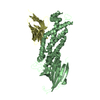

 PDBj
PDBj


 Assembly


 Mass: 35.453 Da / Num. of mol.: 5 / Source method: obtained synthetically / Formula: Cl
 Mass: 106.120 Da / Num. of mol.: 2 / Source method: obtained synthetically / Formula: C4H10O3
 Mass: 18.015 Da / Num. of mol.: 79 / Source method: isolated from a natural source / Formula: H2O
 Sample preparation
 / Beamline: BL11-1 / Wavelength: 0.91162,0.9786,0.97805
 Processing