 Movie
Movie Controller


+ Open data

- Basic information
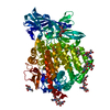
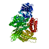
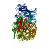
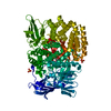
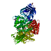
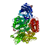
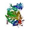
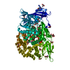
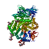
| Entry | Database: PDB / ID: 4kx7 | |||||||||
|---|---|---|---|---|---|---|---|---|---|---|
| Title | Crystal structure of human aminopeptidase A | |||||||||
 Components Components | Glutamyl aminopeptidase | |||||||||
 Keywords Keywords | HYDROLASE / zinc-aminopeptidase fold | |||||||||
| Function / homology |  Function and homology information Function and homology informationglutamyl aminopeptidase / glutamyl aminopeptidase activity / glomerulus development / peptide catabolic process / metalloaminopeptidase activity / brush border / regulation of systemic arterial blood pressure by renin-angiotensin / aminopeptidase activity / Metabolism of Angiotensinogen to Angiotensins / angiotensin maturation ...glutamyl aminopeptidase / glutamyl aminopeptidase activity / glomerulus development / peptide catabolic process / metalloaminopeptidase activity / brush border / regulation of systemic arterial blood pressure by renin-angiotensin / aminopeptidase activity / Metabolism of Angiotensinogen to Angiotensins / angiotensin maturation / apical part of cell / cell-cell signaling / cell migration / angiogenesis / cytoplasmic vesicle / cell population proliferation / apical plasma membrane / external side of plasma membrane / lysosomal membrane / : / extracellular exosome / zinc ion binding / plasma membrane Similarity search - Function | |||||||||
| Biological species |  Homo sapiens (human) Homo sapiens (human) | |||||||||
| Method |  X-RAY DIFFRACTION / SYNCHROTRON / MIR / Resolution: 2.15 Å X-RAY DIFFRACTION / SYNCHROTRON / MIR / Resolution: 2.15 Å | |||||||||
 Authors Authors | Yang, Y. / Liu, C. / Lin, Y.Y. / Li, F. | |||||||||
 Citation Citation | Journal: J.Biol.Chem. / Year: 2013 Title: Structural insights into central hypertension regulation by human aminopeptidase a. Authors: Yang, Y. / Liu, C. / Lin, Y.L. / Li, F. | |||||||||
| History |
|
- Structure visualization
| Structure viewer | Molecule: MolmilJmol/JSmol |
|---|
- Downloads & links
-Download
| PDBx/mmCIF format | 4kx7.cif.gz | 415.4 KB | Display | PDBx/mmCIF format |
|---|---|---|---|---|
| PDB format | pdb4kx7.ent.gz | 340.6 KB | Display | PDB format |
| PDBx/mmJSON format | 4kx7.json.gz | Tree view | PDBx/mmJSON format | |
| Others |  Other downloads Other downloads |
-Validation report
| Arichive directory | https://data.pdbj.org/pub/pdb/validation_reports/kx/4kx7ftp://data.pdbj.org/pub/pdb/validation_reports/kx/4kx7 | HTTPS FTP |
|---|
-Related structure data
| Related structure data |  4kx8C  4kx9C  4kxaC  4kxbC  4kxcC  4kxdC C: citing same article ( |
|---|---|
| Similar structure data |

-Links
 PDBj
PDBj


- Assembly
| Deposited unit | 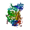
| ||||||||
|---|---|---|---|---|---|---|---|---|---|
| 1 |
| ||||||||
| Unit cell |
|
-Components
-Protein , 1 types, 1 molecules A
| #1: Protein | Mass: 102719.477 Da / Num. of mol.: 1 / Fragment: UNP residues 76-957 Source method: isolated from a genetically manipulated source Source: (gene. exp.) Homo sapiens (human) / Gene: ENPEP / Plasmid: pFastBacTM / Production host:   Spodoptera frugiperda (fall armyworm) / Strain (production host): SF9 / References: UniProt: Q07075, glutamyl aminopeptidase Spodoptera frugiperda (fall armyworm) / Strain (production host): SF9 / References: UniProt: Q07075, glutamyl aminopeptidase |
|---|
-Sugars , 3 types, 8 molecules 
| #2: Polysaccharide | Source method: isolated from a genetically manipulated source #3: Polysaccharide |   Source method: isolated from a genetically manipulated source Details: oligosaccharide / References: triacetyl-beta-chitotriose #5: Sugar |  Type: D-saccharide, beta linking / Mass: 221.208 Da / Num. of mol.: 3 Type: D-saccharide, beta linking / Mass: 221.208 Da / Num. of mol.: 3Source method: isolated from a genetically manipulated source Formula: C8H15NO6 |
|---|
-Non-polymers , 2 types, 595 molecules 

| #4: Chemical | ChemComp-ZN /  Mass: 65.409 Da / Num. of mol.: 1 / Source method: obtained synthetically / Formula: Zn Mass: 65.409 Da / Num. of mol.: 1 / Source method: obtained synthetically / Formula: Zn |
|---|---|
| #6: Water | ChemComp-HOH / Mass: 18.015 Da / Num. of mol.: 594 / Source method: isolated from a natural source / Formula: H2O |
-Details
| Has protein modification | Y |
|---|
-Experimental details
-Experiment
| Experiment | Method: X-RAY DIFFRACTION / Number of used crystals: 1 |
|---|
- Sample preparation
| Crystal | Density Matthews: 3.39 Å3/Da / Density % sol: 63.71 % |
|---|---|
| Crystal grow | Temperature: 277 K / Method: vapor diffusion, sitting drop / pH: 7 Details: 1.6 M sodium citrate pH 7.0, VAPOR DIFFUSION, SITTING DROP, temperature 277K |
-Data collection
| Diffraction source | Source: SYNCHROTRON / Site: ALS  / Beamline: 4.2.2 / Beamline: 4.2.2 |
|---|---|
| Detector | Date: May 17, 2012 |
| Radiation | Protocol: SINGLE WAVELENGTH / Monochromatic (M) / Laue (L): M / Scattering type: x-ray |
| Radiation wavelength | Relative weight: 1 |
| Reflection | Resolution: 2.15→40 Å / Num. obs: 72327 |
- Processing
| Software | Name: REFMAC / Version: 5.7.0029 / Classification: refinement | ||||||||||||||||||||||||||||||||||||||||||||||||||||||||||||||||||||||||||||||||||||||||||||||||||||||||||||||||||||||||||||||||||||||||||||||||||||||||||||||||||||||||||||||||||||||
|---|---|---|---|---|---|---|---|---|---|---|---|---|---|---|---|---|---|---|---|---|---|---|---|---|---|---|---|---|---|---|---|---|---|---|---|---|---|---|---|---|---|---|---|---|---|---|---|---|---|---|---|---|---|---|---|---|---|---|---|---|---|---|---|---|---|---|---|---|---|---|---|---|---|---|---|---|---|---|---|---|---|---|---|---|---|---|---|---|---|---|---|---|---|---|---|---|---|---|---|---|---|---|---|---|---|---|---|---|---|---|---|---|---|---|---|---|---|---|---|---|---|---|---|---|---|---|---|---|---|---|---|---|---|---|---|---|---|---|---|---|---|---|---|---|---|---|---|---|---|---|---|---|---|---|---|---|---|---|---|---|---|---|---|---|---|---|---|---|---|---|---|---|---|---|---|---|---|---|---|---|---|---|---|
| Refinement | Method to determine structure: MIR / Resolution: 2.15→38.86 Å / Cor.coef. Fo:Fc: 0.969 / Cor.coef. Fo:Fc free: 0.949 / SU B: 13.773 / SU ML: 0.146 / Cross valid method: THROUGHOUT / ESU R: 2.895 / ESU R Free: 0.16 / Stereochemistry target values: MAXIMUM LIKELIHOOD
| ||||||||||||||||||||||||||||||||||||||||||||||||||||||||||||||||||||||||||||||||||||||||||||||||||||||||||||||||||||||||||||||||||||||||||||||||||||||||||||||||||||||||||||||||||||||
| Solvent computation | Ion probe radii: 0.8 Å / Shrinkage radii: 0.8 Å / VDW probe radii: 1.4 Å / Solvent model: BABINET MODEL WITH MASK | ||||||||||||||||||||||||||||||||||||||||||||||||||||||||||||||||||||||||||||||||||||||||||||||||||||||||||||||||||||||||||||||||||||||||||||||||||||||||||||||||||||||||||||||||||||||
| Displacement parameters | Biso mean: 58.962 Å2
| ||||||||||||||||||||||||||||||||||||||||||||||||||||||||||||||||||||||||||||||||||||||||||||||||||||||||||||||||||||||||||||||||||||||||||||||||||||||||||||||||||||||||||||||||||||||
| Refinement step | Cycle: LAST / Resolution: 2.15→38.86 Å
| ||||||||||||||||||||||||||||||||||||||||||||||||||||||||||||||||||||||||||||||||||||||||||||||||||||||||||||||||||||||||||||||||||||||||||||||||||||||||||||||||||||||||||||||||||||||
| Refine LS restraints |
|