 Movie
Movie Controller

[English] 日本語
 Yorodumi
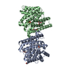
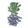
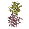
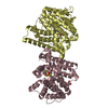
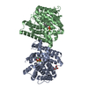
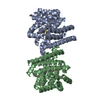
- PDB-4kvi: Crystal structure of Aspergillus terreus aristolochene synthase c... -
+ Open data

- Basic information
| Entry | Database: PDB / ID: 4kvi | ||||||
|---|---|---|---|---|---|---|---|
| Title | Crystal structure of Aspergillus terreus aristolochene synthase complexed with (4aS,7S)-4a-methyl-7-(prop-1-en-2-yl)-2,3,4,4a,5,6,7,8-octahydroquinolin-1-ium | ||||||
 Components Components | Aristolochene synthase | ||||||
 Keywords Keywords | LYASE/LYASE INHIBITOR / Class I terpene cyclase / alpha-helical fold / farnesyl diphosphate / metal-binding / magnesium / LYASE-LYASE INHIBITOR complex | ||||||
| Function / homology |  Function and homology information Function and homology informationaristolochene synthase / aristolochene synthase activity / isoprenoid biosynthetic process / metal ion binding Similarity search - Function | ||||||
| Biological species |  | ||||||
| Method |  X-RAY DIFFRACTION / SYNCHROTRON / MOLECULAR REPLACEMENT / Resolution: 2.15 Å X-RAY DIFFRACTION / SYNCHROTRON / MOLECULAR REPLACEMENT / Resolution: 2.15 Å | ||||||
 Authors Authors | Chen, M. / Faraldos, J.A. / Al-lami, N. / Janvier, M. / D'Antonio, E.L. / Cane, D.E. / Allemann, R.K. / Christianson, D.W. | ||||||
 Citation Citation | Journal: Biochemistry / Year: 2013 Title: Mechanistic insights from the binding of substrate and carbocation intermediate analogues to aristolochene synthase. Authors: Chen, M. / Al-Lami, N. / Janvier, M. / D'Antonio, E.L. / Faraldos, J.A. / Cane, D.E. / Allemann, R.K. / Christianson, D.W. | ||||||
| History |
|
- Structure visualization
| Structure viewer | Molecule: MolmilJmol/JSmol |
|---|
- Downloads & links
-Download
| PDBx/mmCIF format | 4kvi.cif.gz | 275.5 KB | Display | PDBx/mmCIF format |
|---|---|---|---|---|
| PDB format | pdb4kvi.ent.gz | 221.1 KB | Display | PDB format |
| PDBx/mmJSON format | 4kvi.json.gz | Tree view | PDBx/mmJSON format | |
| Others |  Other downloads Other downloads |
-Validation report
| Arichive directory | https://data.pdbj.org/pub/pdb/validation_reports/kv/4kviftp://data.pdbj.org/pub/pdb/validation_reports/kv/4kvi | HTTPS FTP |
|---|
-Related structure data
| Related structure data |  4kuxC 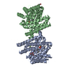 4kvdSC 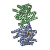 4kvwC  4kvyC 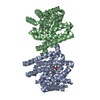 4kwdC C: citing same article ( S: Starting model for refinement |
|---|---|
| Similar structure data |


-Links
 PDBj
PDBj


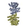
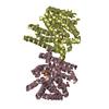
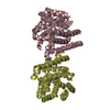
- Assembly
| Deposited unit | 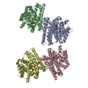
| ||||||||
|---|---|---|---|---|---|---|---|---|---|
| 1 | 
| ||||||||
| 2 | 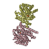
| ||||||||
| Unit cell |
|
-Components
| #1: Protein | Mass: 36221.395 Da / Num. of mol.: 4 / Fragment: UNP residues 14-320 Source method: isolated from a genetically manipulated source Source: (gene. exp.)  #2: Chemical | ChemComp-MG /   Mass: 24.305 Da / Num. of mol.: 12 / Source method: obtained synthetically / Formula: Mg Mass: 24.305 Da / Num. of mol.: 12 / Source method: obtained synthetically / Formula: Mg#3: Chemical | ChemComp-POP /   Mass: 175.959 Da / Num. of mol.: 4 / Source method: obtained synthetically / Formula: H2O7P2 Mass: 175.959 Da / Num. of mol.: 4 / Source method: obtained synthetically / Formula: H2O7P2#4: Chemical | ChemComp-1SV / ( 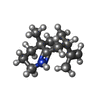  Mass: 192.320 Da / Num. of mol.: 4 / Source method: obtained synthetically / Formula: C13H22N Mass: 192.320 Da / Num. of mol.: 4 / Source method: obtained synthetically / Formula: C13H22N#5: Water | ChemComp-HOH / |  Mass: 18.015 Da / Num. of mol.: 892 / Source method: isolated from a natural source / Formula: H2O Mass: 18.015 Da / Num. of mol.: 892 / Source method: isolated from a natural source / Formula: H2O |
|---|
-Experimental details
-Experiment
| Experiment | Method: X-RAY DIFFRACTION / Number of used crystals: 1 |
|---|
- Sample preparation
| Crystal | Density Matthews: 3.06 Å3/Da / Density % sol: 59.78 % |
|---|---|
| Crystal grow | Temperature: 277 K / Method: vapor diffusion, hanging drop / pH: 7.7 Details: 3 uL protein (10 mg/mL ATAS, 20 mM MES, pH 6.5, 1.9 mM magnesium chloride, 120 mM sodium chloride, 3 mM BME, 1.6 mM sodium pyrophosphate, 1.5 mM inhibitor) + 3 uL precipitant (100 mM HEPES, ...Details: 3 uL protein (10 mg/mL ATAS, 20 mM MES, pH 6.5, 1.9 mM magnesium chloride, 120 mM sodium chloride, 3 mM BME, 1.6 mM sodium pyrophosphate, 1.5 mM inhibitor) + 3 uL precipitant (100 mM HEPES, pH 7.7, 200 mM magnesium chloride, 23% w/v PEG3350) against 500 uL precipitant, VAPOR DIFFUSION, HANGING DROP, temperature 277K |
-Data collection
| Diffraction | Mean temperature: 100 K |
|---|---|
| Diffraction source | Source: SYNCHROTRON / Site: NSLS  / Beamline: X29A / Wavelength: 1.075 Å / Beamline: X29A / Wavelength: 1.075 Å |
| Detector | Type: ADSC QUANTUM 315r / Detector: CCD / Date: Jul 25, 2012 Details: Cryogenically cooled double crystal monochromator with horizontally focusing sagitally bent second crystal with 4:1 magnification ratio and vertically focusing mirror |
| Radiation | Monochromator: Sagitally focused Si(111) / Protocol: SINGLE WAVELENGTH / Monochromatic (M) / Laue (L): M / Scattering type: x-ray |
| Radiation wavelength | Wavelength: 1.075 Å / Relative weight: 1 |
| Reflection | Resolution: 2.15→50 Å / Num. obs: 96118 / % possible obs: 99.1 % / Observed criterion σ(F): 0 / Observed criterion σ(I): -3 / Redundancy: 5.6 % / Rmerge(I) obs: 0.106 / Rsym value: 0.106 / Net I/σ(I): 16.304 |
| Reflection shell | Resolution: 2.15→2.23 Å / Redundancy: 5.6 % / Rmerge(I) obs: 0.569 / Mean I/σ(I) obs: 3.25 / Rsym value: 0.569 / % possible all: 99.9 |
- Processing
| Software |
| |||||||||||||||||||||||||||||||||||||||||||||||||||||||||||||||||||||||||||||||||||||||||||||||||||||||||||||||||||||||||||||||||||||||||||||||||||||||||||||||||||||||||||||||||||||||||||||||||||||||||||||||||||||||||
|---|---|---|---|---|---|---|---|---|---|---|---|---|---|---|---|---|---|---|---|---|---|---|---|---|---|---|---|---|---|---|---|---|---|---|---|---|---|---|---|---|---|---|---|---|---|---|---|---|---|---|---|---|---|---|---|---|---|---|---|---|---|---|---|---|---|---|---|---|---|---|---|---|---|---|---|---|---|---|---|---|---|---|---|---|---|---|---|---|---|---|---|---|---|---|---|---|---|---|---|---|---|---|---|---|---|---|---|---|---|---|---|---|---|---|---|---|---|---|---|---|---|---|---|---|---|---|---|---|---|---|---|---|---|---|---|---|---|---|---|---|---|---|---|---|---|---|---|---|---|---|---|---|---|---|---|---|---|---|---|---|---|---|---|---|---|---|---|---|---|---|---|---|---|---|---|---|---|---|---|---|---|---|---|---|---|---|---|---|---|---|---|---|---|---|---|---|---|---|---|---|---|---|---|---|---|---|---|---|---|---|---|---|---|---|---|---|---|---|
| Refinement | Method to determine structure: MOLECULAR REPLACEMENT Starting model: PDB ENTRY 4KVD Resolution: 2.15→50 Å / SU ML: 0.22 / Isotropic thermal model: ISOTROPIC / Cross valid method: THROUGHOUT / σ(F): 1.34 / Phase error: 23.71 / Stereochemistry target values: Engh & Huber
| |||||||||||||||||||||||||||||||||||||||||||||||||||||||||||||||||||||||||||||||||||||||||||||||||||||||||||||||||||||||||||||||||||||||||||||||||||||||||||||||||||||||||||||||||||||||||||||||||||||||||||||||||||||||||
| Solvent computation | Shrinkage radii: 0.9 Å / VDW probe radii: 1.11 Å / Solvent model: FLAT BULK SOLVENT MODEL | |||||||||||||||||||||||||||||||||||||||||||||||||||||||||||||||||||||||||||||||||||||||||||||||||||||||||||||||||||||||||||||||||||||||||||||||||||||||||||||||||||||||||||||||||||||||||||||||||||||||||||||||||||||||||
| Displacement parameters | Biso mean: 31 Å2 | |||||||||||||||||||||||||||||||||||||||||||||||||||||||||||||||||||||||||||||||||||||||||||||||||||||||||||||||||||||||||||||||||||||||||||||||||||||||||||||||||||||||||||||||||||||||||||||||||||||||||||||||||||||||||
| Refine analyze |
| |||||||||||||||||||||||||||||||||||||||||||||||||||||||||||||||||||||||||||||||||||||||||||||||||||||||||||||||||||||||||||||||||||||||||||||||||||||||||||||||||||||||||||||||||||||||||||||||||||||||||||||||||||||||||
| Refinement step | Cycle: LAST / Resolution: 2.15→50 Å
| |||||||||||||||||||||||||||||||||||||||||||||||||||||||||||||||||||||||||||||||||||||||||||||||||||||||||||||||||||||||||||||||||||||||||||||||||||||||||||||||||||||||||||||||||||||||||||||||||||||||||||||||||||||||||
| Refine LS restraints |
| |||||||||||||||||||||||||||||||||||||||||||||||||||||||||||||||||||||||||||||||||||||||||||||||||||||||||||||||||||||||||||||||||||||||||||||||||||||||||||||||||||||||||||||||||||||||||||||||||||||||||||||||||||||||||
| LS refinement shell |
|
