 Movie
Movie Controller

[English] 日本語
 Yorodumi
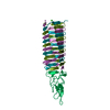
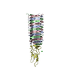
- PDB-4ku0: Enterobacteria phage T4 gp5.4 PAAR repeat protein in complex with... -
+ Open data

- Basic information
| Entry | Database: PDB / ID: 4ku0 | ||||||
|---|---|---|---|---|---|---|---|
| Title | Enterobacteria phage T4 gp5.4 PAAR repeat protein in complex with T4 gp5 beta-helix fragment | ||||||
 Components Components |
| ||||||
 Keywords Keywords | HYDROLASE/PROTEIN BINDING / PAAR-repeat motif / membrane piercing / type VI secretion system / T6SS / cell puncturing device / beta-helix / gp5-gp27 protein complex / HYDROLASE-PROTEIN BINDING complex | ||||||
| Function / homology |  Function and homology information Function and homology informationpeptidoglycan beta-N-acetylmuramidase activity / symbiont entry into host cell via disruption of host cell wall peptidoglycan / virus tail, baseplate / viral tail assembly / symbiont entry into host cell via disruption of host cell envelope / symbiont entry into host / virus tail / peptidoglycan catabolic process / cell wall macromolecule catabolic process / lysozyme ...peptidoglycan beta-N-acetylmuramidase activity / symbiont entry into host cell via disruption of host cell wall peptidoglycan / virus tail, baseplate / viral tail assembly / symbiont entry into host cell via disruption of host cell envelope / symbiont entry into host / virus tail / peptidoglycan catabolic process / cell wall macromolecule catabolic process / lysozyme / lysozyme activity / killing of cells of another organism / defense response to bacterium / symbiont entry into host cell / metal ion binding / identical protein binding Similarity search - Function | ||||||
| Biological species |  Enterobacteria phage T4 (virus) Enterobacteria phage T4 (virus) | ||||||
| Method |  X-RAY DIFFRACTION / SYNCHROTRON / MOLECULAR REPLACEMENT / Resolution: 1.15 Å X-RAY DIFFRACTION / SYNCHROTRON / MOLECULAR REPLACEMENT / Resolution: 1.15 Å | ||||||
 Authors Authors | Buth, S.A. / Leiman, P.G. / Shneider, M.M. | ||||||
 Citation Citation | Journal: To be Published Title: Crystall structute of the business end of the T4 cell-puncturing device Authors: Buth, S.A. / Leiman, P.G. / Shneider, M.M. | ||||||
| History |
|
- Structure visualization
| Structure viewer | Molecule: MolmilJmol/JSmol |
|---|
- Downloads & links
-Download
| PDBx/mmCIF format | 4ku0.cif.gz | 187.9 KB | Display | PDBx/mmCIF format |
|---|---|---|---|---|
| PDB format | pdb4ku0.ent.gz | 148.4 KB | Display | PDB format |
| PDBx/mmJSON format | 4ku0.json.gz | Tree view | PDBx/mmJSON format | |
| Others |  Other downloads Other downloads |
-Validation report
| Arichive directory | https://data.pdbj.org/pub/pdb/validation_reports/ku/4ku0ftp://data.pdbj.org/pub/pdb/validation_reports/ku/4ku0 | HTTPS FTP |
|---|
-Related structure data
| Related structure data |  4jj2S S: Starting model for refinement |
|---|---|
| Similar structure data |


-Links
 PDBj
PDBj


- Assembly
| Deposited unit | 
| ||||||||
|---|---|---|---|---|---|---|---|---|---|
| 1 |
| ||||||||
| Unit cell |
|
-Components
-Protein , 2 types, 4 molecules ABCD
| #1: Protein | Mass: 9856.678 Da / Num. of mol.: 3 / Fragment: residues 484-575 Source method: isolated from a genetically manipulated source Source: (gene. exp.) Enterobacteria phage T4 (virus) / Gene: 5 / Plasmid: pEEva2, a pET23a derivative / Production host:  #2: Protein | | Mass: 10102.467 Da / Num. of mol.: 1 Source method: isolated from a genetically manipulated source Source: (gene. exp.) Enterobacteria phage T4 (virus) / Gene: 5.4, y08B / Plasmid: pEEva2, a pET23a derivative / Production host: |
|---|
-Non-polymers , 8 types, 571 molecules 


| #3: Chemical | ChemComp-MG /  Mass: 24.305 Da / Num. of mol.: 1 / Source method: obtained synthetically / Formula: Mg Mass: 24.305 Da / Num. of mol.: 1 / Source method: obtained synthetically / Formula: Mg | ||||||||||
|---|---|---|---|---|---|---|---|---|---|---|---|
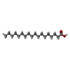
| #4: Chemical | ChemComp-ELA /  Mass: 282.461 Da / Num. of mol.: 1 / Source method: obtained synthetically / Formula: C18H34O2 Mass: 282.461 Da / Num. of mol.: 1 / Source method: obtained synthetically / Formula: C18H34O2 | ||||||||||
| #5: Chemical | ChemComp-EDO /  Mass: 62.068 Da / Num. of mol.: 4 / Source method: obtained synthetically / Formula: C2H6O2 Mass: 62.068 Da / Num. of mol.: 4 / Source method: obtained synthetically / Formula: C2H6O2#6: Chemical | ChemComp-STE / |  Mass: 284.477 Da / Num. of mol.: 1 / Source method: obtained synthetically / Formula: C18H36O2 Mass: 284.477 Da / Num. of mol.: 1 / Source method: obtained synthetically / Formula: C18H36O2#7: Chemical | ChemComp-PLM / |  Mass: 256.424 Da / Num. of mol.: 1 / Source method: obtained synthetically / Formula: C16H32O2 Mass: 256.424 Da / Num. of mol.: 1 / Source method: obtained synthetically / Formula: C16H32O2#8: Chemical | ChemComp-FE / |  Mass: 55.845 Da / Num. of mol.: 1 / Source method: obtained synthetically / Formula: Fe Mass: 55.845 Da / Num. of mol.: 1 / Source method: obtained synthetically / Formula: Fe#9: Chemical | ChemComp-NA / |  Mass: 22.990 Da / Num. of mol.: 1 / Source method: obtained synthetically / Formula: Na Mass: 22.990 Da / Num. of mol.: 1 / Source method: obtained synthetically / Formula: Na#10: Water | ChemComp-HOH / | Mass: 18.015 Da / Num. of mol.: 561 / Source method: isolated from a natural source / Formula: H2O |
-Details
| Has protein modification | N |
|---|
-Experimental details
-Experiment
| Experiment | Method: X-RAY DIFFRACTION / Number of used crystals: 1 |
|---|
- Sample preparation
| Crystal | Density Matthews: 2.41 Å3/Da / Density % sol: 48.87 % |
|---|---|
| Crystal grow | Temperature: 293 K / Method: vapor diffusion, hanging drop / pH: 8.5 Details: 23-25% PEG 3350, 100mM Tris pH=8.5, 40-100mM MgCl2, VAPOR DIFFUSION, HANGING DROP, temperature 293K |
-Data collection
| Diffraction | Mean temperature: 100 K |
|---|---|
| Diffraction source | Source: SYNCHROTRON / Site: SLS  / Beamline: X06SA / Wavelength: 1 Å / Beamline: X06SA / Wavelength: 1 Å |
| Detector | Type: DECTRIS PILATUS 6M / Detector: PIXEL / Date: Sep 3, 2012 / Details: dynamically bendable mirror |
| Radiation | Monochromator: Si(111) monochromator / Protocol: SINGLE WAVELENGTH / Monochromatic (M) / Laue (L): M / Scattering type: x-ray |
| Radiation wavelength | Wavelength: 1 Å / Relative weight: 1 |
| Reflection | Resolution: 1.15→46 Å / Num. all: 129006 / Num. obs: 124490 / % possible obs: 96.5 % / Observed criterion σ(F): 0 / Observed criterion σ(I): 2 / Redundancy: 6.13 % / Biso Wilson estimate: 18.103 Å2 / Rmerge(I) obs: 0.0532 / Net I/σ(I): 12.12 |
| Reflection shell | Resolution: 1.15→1.22 Å / Redundancy: 4.12 % / Mean I/σ(I) obs: 2.27 / Num. unique all: 21326 / % possible all: 81.3 |
- Processing
| Software |
| |||||||||||||||||||||||||||||||||
|---|---|---|---|---|---|---|---|---|---|---|---|---|---|---|---|---|---|---|---|---|---|---|---|---|---|---|---|---|---|---|---|---|---|---|
| Refinement | Method to determine structure: MOLECULAR REPLACEMENT Starting model: 4JJ2 Resolution: 1.15→46 Å / Num. parameters: 31827 / Num. restraintsaints: 40407 / Cross valid method: THROUGHOUT / σ(F): 4 / Stereochemistry target values: ENGH & HUBER
| |||||||||||||||||||||||||||||||||
| Refine analyze | Num. disordered residues: 23 / Occupancy sum hydrogen: 0 / Occupancy sum non hydrogen: 3356.94 | |||||||||||||||||||||||||||||||||
| Refinement step | Cycle: LAST / Resolution: 1.15→46 Å
| |||||||||||||||||||||||||||||||||
| Refine LS restraints |
|
