Resolution: 2.2→2.28 Å / Redundancy: 16.6 % / Rmerge(I) obs: 0.628 / Mean I/σ(I) obs: 5.256 / Num. unique all: 2551 / Rsym value: 0.628 / % possible all: 100
-
Processing
Software
Name
Version
Classification
CrystalClear
datacollection
PHASER
phasing
REFMAC
5.6.0117
refinement
HKL-2000
datareduction
HKL-2000
datascaling
Refinement
Method to determine structure: MOLECULAR REPLACEMENT / Resolution: 2.2→29.14 Å / Cor.coef. Fo:Fc: 0.968 / Cor.coef. Fo:Fc free: 0.951 / SU B: 5.747 / SU ML: 0.078 / Cross valid method: THROUGHOUT / σ(I): 5 / ESU R: 0.145 / ESU R Free: 0.134 / Stereochemistry target values: MAXIMUM LIKELIHOOD / Details: HYDROGENS HAVE BEEN USED IF PRESENT IN THE INPUT
Rfactor
Num. reflection
% reflection
Selection details
Rfree
0.1735
2033
5 %
RANDOM
Rwork
0.13674
-
-
-
obs
0.13854
38529
99.9 %
-
all
-
40562
-
-
Solvent computation
Ion probe radii: 0.8 Å / Shrinkage radii: 0.8 Å / VDW probe radii: 1.2 Å / Solvent model: MASK
Displacement parameters
Biso mean: 24.377 Å2
Baniso -1
Baniso -2
Baniso -3
1-
-0.05 Å2
-0.02 Å2
0 Å2
2-
-
-0.05 Å2
0 Å2
3-
-
-
0.07 Å2
Refinement step
Cycle: LAST / Resolution: 2.2→29.14 Å
Protein
Nucleic acid
Ligand
Solvent
Total
Num. atoms
3782
0
58
472
4312
Refine LS restraints
Refine-ID
Type
Dev ideal
Dev ideal target
Number
X-RAY DIFFRACTION
r_bond_refined_d
0.017
0.019
4182
X-RAY DIFFRACTION
r_angle_refined_deg
1.749
1.978
5728
X-RAY DIFFRACTION
r_dihedral_angle_1_deg
6.061
5
602
X-RAY DIFFRACTION
r_dihedral_angle_2_deg
39.297
25.647
170
X-RAY DIFFRACTION
r_dihedral_angle_3_deg
14.304
15
724
X-RAY DIFFRACTION
r_dihedral_angle_4_deg
16.665
15
21
X-RAY DIFFRACTION
r_chiral_restr
0.115
0.2
669
X-RAY DIFFRACTION
r_gen_planes_refined
0.009
0.021
3176
LS refinement shell
Resolution: 2.2→2.257 Å / Total num. of bins used: 20
Rfactor
Num. reflection
% reflection
Rfree
0.217
120
-
Rwork
0.154
2551
-
obs
-
2551
100 %
Refinement TLS params.
Method: refined / Refine-ID: X-RAY DIFFRACTION
ID
L11 (°2)
L12 (°2)
L13 (°2)
L22 (°2)
L23 (°2)
L33 (°2)
S11 (Å °)
S12 (Å °)
S13 (Å °)
S21 (Å °)
S22 (Å °)
S23 (Å °)
S31 (Å °)
S32 (Å °)
S33 (Å °)
T11 (Å2)
T12 (Å2)
T13 (Å2)
T22 (Å2)
T23 (Å2)
T33 (Å2)
Origin x (Å)
Origin y (Å)
Origin z (Å)
1
1.5532
-0.6825
-0.1409
0.5918
-0.0993
0.4115
-0.0375
-0.0995
-0.2375
0.0765
0.1366
0.162
-0.0534
-0.1071
-0.0991
0.1043
-0.0061
-0.0036
0.1115
0.0237
0.1112
32.286
-61.923
22.187
2
0.7091
0.2669
-0.3213
0.5883
-0.2693
0.5005
-0.0134
-0.0319
-0.0009
-0.0031
0.0267
0.0321
0.0311
0.0135
-0.0133
0.0544
-0.0025
-0.03
0.0349
-0.009
0.0258
39.238
-52.627
10.693
3
0.4372
0.1339
-0.005
0.2714
0.0406
0.2979
-0.0215
0.0209
0.0438
-0.026
-0.004
0.0352
-0.0169
-0.007
0.0255
0.0443
0.0048
-0.0222
0.0315
-0.0008
0.0159
57.653
-33.741
6.845
Refinement TLS group
ID
Refine-ID
Refine TLS-ID
Auth asym-ID
Auth seq-ID
1
X-RAY DIFFRACTION
1
A
55 - 145
2
X-RAY DIFFRACTION
2
A
146 - 284
3
X-RAY DIFFRACTION
3
A
285 - 571
+
About Yorodumi
-
News
-
Feb 9, 2022. New format data for meta-information of EMDB entries
New format data for meta-information of EMDB entries
Version 3 of the EMDB header file is now the official format.
The previous official version 1.9 will be removed from the archive.
In the structure databanks used in Yorodumi, some data are registered as the other names, "COVID-19 virus" and "2019-nCoV". Here are the details of the virus and the list of structure data.
Jan 31, 2019. EMDB accession codes are about to change! (news from PDBe EMDB page)
EMDB accession codes are about to change! (news from PDBe EMDB page)
The allocation of 4 digits for EMDB accession codes will soon come to an end. Whilst these codes will remain in use, new EMDB accession codes will include an additional digit and will expand incrementally as the available range of codes is exhausted. The current 4-digit format prefixed with “EMD-” (i.e. EMD-XXXX) will advance to a 5-digit format (i.e. EMD-XXXXX), and so on. It is currently estimated that the 4-digit codes will be depleted around Spring 2019, at which point the 5-digit format will come into force.
The EM Navigator/Yorodumi systems omit the EMD- prefix.
Related info.:Q: What is EMD? / ID/Accession-code notation in Yorodumi/EM Navigator
Yorodumi is a browser for structure data from EMDB, PDB, SASBDB, etc.
This page is also the successor to EM Navigator detail page, and also detail information page/front-end page for Omokage search.
The word "yorodu" (or yorozu) is an old Japanese word meaning "ten thousand". "mi" (miru) is to see.
Related info.:EMDB / PDB / SASBDB / Comparison of 3 databanks / Yorodumi Search / Aug 31, 2016. New EM Navigator & Yorodumi / Yorodumi Papers / Jmol/JSmol / Function and homology information / Changes in new EM Navigator and Yorodumi
 Movie
Movie Controller

 Yorodumi
 Open data
 Basic information
 Components
 Keywords
 Function and homology information
 X-RAY DIFFRACTION /
  Authors
 Citation
 Structure visualization
 Downloads & links
 Other downloads
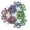


 PDBj
PDBj


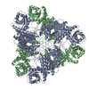
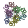
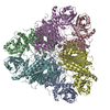
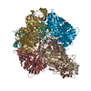
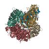
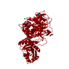
 Assembly


 Mass: 24.305 Da / Num. of mol.: 2 / Source method: obtained synthetically / Formula: Mg
 Mass: 96.063 Da / Num. of mol.: 2 / Source method: obtained synthetically / Formula: SO4
 Mass: 35.453 Da / Num. of mol.: 3 / Source method: obtained synthetically / Formula: Cl
 Mass: 92.094 Da / Num. of mol.: 5 / Source method: obtained synthetically / Formula: C3H8O3
 Mass: 69.085 Da / Num. of mol.: 1 / Source method: obtained synthetically / Formula: C3H5N2
 Mass: 118.174 Da / Num. of mol.: 1 / Source method: obtained synthetically / Formula: C6H14O2 / Comment: precipitant*YM
 Sample preparation
 Processing