 Movie
Movie Controller

[English] 日本語
 Yorodumi
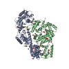
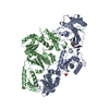
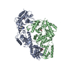
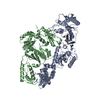
- PDB-4ko0: CRYSTAL STRUCTURE OF HIV-1 REVERSE TRANSCRIPTASE (RT) IN COMPLEX ... -
+ Open data

- Basic information
| Entry | Database: PDB / ID: 4ko0 | ||||||
|---|---|---|---|---|---|---|---|
| Title | CRYSTAL STRUCTURE OF HIV-1 REVERSE TRANSCRIPTASE (RT) IN COMPLEX WITH an anilinylpyrimidine derivative (JLJ-135) | ||||||
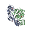
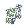
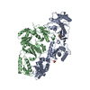
 Components Components | (HIV-1 reverse transcriptase, ...) x 2 | ||||||
 Keywords Keywords | HYDROLASE/HYDROLASE INHIBITOR / P51/P66 / HETERO DIMER / NNRTI / NONNUCLEOSIDE INHIBITOR / AIDS / HIV / DNA RECOMBINATION / RNA-DIRECTED DNA POLYMERASE / DNA POLYMERASE / ENDONUCLEASE / HYDROLASE / MULTIFUNCTIONAL ENZYME / TRANSFERASE / HYDROLASE-INHIBITOR COMPLEX / HYDROLASE-HYDROLASE INHIBITOR complex | ||||||
| Function / homology |  Function and homology information Function and homology informationHIV-1 retropepsin / symbiont-mediated activation of host apoptosis / retroviral ribonuclease H / exoribonuclease H / exoribonuclease H activity / host multivesicular body / DNA integration / viral genome integration into host DNA / RNA-directed DNA polymerase / establishment of integrated proviral latency ...HIV-1 retropepsin / symbiont-mediated activation of host apoptosis / retroviral ribonuclease H / exoribonuclease H / exoribonuclease H activity / host multivesicular body / DNA integration / viral genome integration into host DNA / RNA-directed DNA polymerase / establishment of integrated proviral latency / symbiont-mediated suppression of host gene expression / viral penetration into host nucleus / RNA stem-loop binding / RNA-directed DNA polymerase activity / RNA-DNA hybrid ribonuclease activity / Transferases; Transferring phosphorus-containing groups; Nucleotidyltransferases / host cell / viral nucleocapsid / DNA recombination / DNA-directed DNA polymerase / Hydrolases; Acting on ester bonds / aspartic-type endopeptidase activity / DNA-directed DNA polymerase activity / viral translational frameshifting / symbiont entry into host cell / lipid binding / host cell nucleus / host cell plasma membrane / structural molecule activity / virion membrane / proteolysis / DNA binding / zinc ion binding / membrane Similarity search - Function | ||||||
| Biological species |   Human immunodeficiency virus type 1 Human immunodeficiency virus type 1 | ||||||
| Method |  X-RAY DIFFRACTION / SYNCHROTRON / MOLECULAR REPLACEMENT / Resolution: 1.95 Å X-RAY DIFFRACTION / SYNCHROTRON / MOLECULAR REPLACEMENT / Resolution: 1.95 Å | ||||||
 Authors Authors | Das, K. / Bauman, J.D. / Arnold, E. | ||||||
 Citation Citation | Journal: Bioorg.Med.Chem.Lett. / Year: 2013 Title: Extension into the entrance channel of HIV-1 reverse transcriptase-Crystallography and enhanced solubility. Authors: Bollini, M. / Frey, K.M. / Cisneros, J.A. / Spasov, K.A. / Das, K. / Bauman, J.D. / Arnold, E. / Anderson, K.S. / Jorgensen, W.L. | ||||||
| History |
|
- Structure visualization
| Structure viewer | Molecule: MolmilJmol/JSmol |
|---|
- Downloads & links
-Download
| PDBx/mmCIF format | 4ko0.cif.gz | 217.7 KB | Display | PDBx/mmCIF format |
|---|---|---|---|---|
| PDB format | pdb4ko0.ent.gz | 171.2 KB | Display | PDB format |
| PDBx/mmJSON format | 4ko0.json.gz | Tree view | PDBx/mmJSON format | |
| Others |  Other downloads Other downloads |
-Validation report
| Summary document | 4ko0_validation.pdf.gz | 794.7 KB | Display | wwPDB validaton report |
|---|---|---|---|---|
| Full document | 4ko0_full_validation.pdf.gz | 813.6 KB | Display | |
| Data in XML | 4ko0_validation.xml.gz | 38.8 KB | Display | |
| Data in CIF | 4ko0_validation.cif.gz | 55.1 KB | Display | |
| Arichive directory | https://data.pdbj.org/pub/pdb/validation_reports/ko/4ko0ftp://data.pdbj.org/pub/pdb/validation_reports/ko/4ko0 | HTTPS FTP |
-Related structure data
| Related structure data | 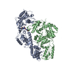 4kkoC  1s9eS C: citing same article ( S: Starting model for refinement |
|---|---|
| Similar structure data |


-Links
 PDBj
PDBj


- Assembly
| Deposited unit | 
| ||||||||
|---|---|---|---|---|---|---|---|---|---|
| 1 |
| ||||||||
| Unit cell |
|
-Components
-HIV-1 reverse transcriptase, ... , 2 types, 2 molecules AB
| #1: Protein | Mass: 63989.238 Da / Num. of mol.: 1 / Fragment: p66 / Mutation: K172A, K173A, C280S Source method: isolated from a genetically manipulated source Source: (gene. exp.) Human immunodeficiency virus type 1 / Strain: 1B1 / Gene: gag-pol / Plasmid: PRT52A / Production host:  |
|---|---|
| #2: Protein | Mass: 50039.488 Da / Num. of mol.: 1 / Fragment: p51 / Mutation: C280S Source method: isolated from a genetically manipulated source Source: (gene. exp.) Human immunodeficiency virus type 1 / Strain: 1B1 / Gene: gag-pol / Plasmid: PRT52A / Production host: |
-Non-polymers , 4 types, 343 molecules 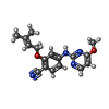


| #3: Chemical | ChemComp-JLJ /  Mass: 310.350 Da / Num. of mol.: 1 / Source method: obtained synthetically / Formula: C17H18N4O2 Mass: 310.350 Da / Num. of mol.: 1 / Source method: obtained synthetically / Formula: C17H18N4O2 | ||||
|---|---|---|---|---|---|
| #4: Chemical |  Mass: 96.063 Da / Num. of mol.: 2 / Source method: obtained synthetically / Formula: SO4 Mass: 96.063 Da / Num. of mol.: 2 / Source method: obtained synthetically / Formula: SO4#5: Chemical |  Mass: 62.068 Da / Num. of mol.: 2 / Source method: obtained synthetically / Formula: C2H6O2 Mass: 62.068 Da / Num. of mol.: 2 / Source method: obtained synthetically / Formula: C2H6O2#6: Water | ChemComp-HOH / | Mass: 18.015 Da / Num. of mol.: 338 / Source method: isolated from a natural source / Formula: H2O |
-Experimental details
-Experiment
| Experiment | Method: X-RAY DIFFRACTION / Number of used crystals: 1 |
|---|
- Sample preparation
| Crystal | Density Matthews: 2.83 Å3/Da / Density % sol: 56.48 % |
|---|---|
| Crystal grow | Temperature: 277 K / pH: 6.8 Details: PEG 8000, AMMONIUM SULFATE, MGCL2, IMIDAZOLE , pH 6.8, VAPOR DIFFUSION, SITTING DROP, temperature 277K |
-Data collection
| Diffraction | Mean temperature: 100 K |
|---|---|
| Diffraction source | Source: SYNCHROTRON / Site: CHESS  / Beamline: F1 / Wavelength: 0.918 / Beamline: F1 / Wavelength: 0.918 |
| Detector | Type: ADSC QUANTUM 4 / Detector: CCD / Date: Jun 9, 2006 / Details: MONOCHROMATOR |
| Radiation | Monochromator: GRAPHITE / Protocol: SINGLE WAVELENGTH / Monochromatic (M) / Laue (L): M / Scattering type: x-ray |
| Radiation wavelength | Wavelength: 0.918 Å / Relative weight: 1 |
| Reflection | Resolution: 1.95→50 Å / Num. obs: 91737 / % possible obs: 98.8 % / Observed criterion σ(I): -1 / Redundancy: 3.6 % / Rmerge(I) obs: 0.058 / Net I/σ(I): 12.3 |
| Reflection shell | Resolution: 1.95→1.98 Å / Redundancy: 2.2 % / Rmerge(I) obs: 0.825 / Mean I/σ(I) obs: 1.3 / % possible all: 99.1 |
- Processing
| Software |
| ||||||||||||||||||||||||||||||||||||||||||||||||||||||||||||||||||||||||||||||||||||||||||||||||||
|---|---|---|---|---|---|---|---|---|---|---|---|---|---|---|---|---|---|---|---|---|---|---|---|---|---|---|---|---|---|---|---|---|---|---|---|---|---|---|---|---|---|---|---|---|---|---|---|---|---|---|---|---|---|---|---|---|---|---|---|---|---|---|---|---|---|---|---|---|---|---|---|---|---|---|---|---|---|---|---|---|---|---|---|---|---|---|---|---|---|---|---|---|---|---|---|---|---|---|---|
| Refinement | Method to determine structure: MOLECULAR REPLACEMENT Starting model: 1S9E Resolution: 1.95→29.31 Å / SU ML: 0.27 / σ(F): 0 / Phase error: 29.78 / Stereochemistry target values: ML
| ||||||||||||||||||||||||||||||||||||||||||||||||||||||||||||||||||||||||||||||||||||||||||||||||||
| Solvent computation | Shrinkage radii: 0.86 Å / VDW probe radii: 1.1 Å / Solvent model: FLAT BULK SOLVENT MODEL / Bsol: 52.01 Å2 / ksol: 0.4 e/Å3 | ||||||||||||||||||||||||||||||||||||||||||||||||||||||||||||||||||||||||||||||||||||||||||||||||||
| Displacement parameters |
| ||||||||||||||||||||||||||||||||||||||||||||||||||||||||||||||||||||||||||||||||||||||||||||||||||
| Refinement step | Cycle: LAST / Resolution: 1.95→29.31 Å
| ||||||||||||||||||||||||||||||||||||||||||||||||||||||||||||||||||||||||||||||||||||||||||||||||||
| Refine LS restraints |
| ||||||||||||||||||||||||||||||||||||||||||||||||||||||||||||||||||||||||||||||||||||||||||||||||||
| LS refinement shell |
|
