 Movie
Movie Controller


+ Open data

- Basic information
| Entry | Database: PDB / ID: 4jw0 | ||||||
|---|---|---|---|---|---|---|---|
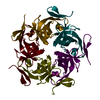
| Title | Structure of Gloeobacter violaceus CcmL | ||||||
 Components Components | Carbon dioxide concentrating mechanism protein | ||||||
 Keywords Keywords | STRUCTURAL PROTEIN / OB fold | ||||||
| Function / homology |  Function and homology information Function and homology informationstructural constituent of carboxysome shell / carboxysome / carbon fixation / photosynthesis Similarity search - Function | ||||||
| Biological species |  Gloeobacter violaceus (bacteria) Gloeobacter violaceus (bacteria) | ||||||
| Method |  X-RAY DIFFRACTION / SYNCHROTRON / MOLECULAR REPLACEMENT / Resolution: 1.7 Å X-RAY DIFFRACTION / SYNCHROTRON / MOLECULAR REPLACEMENT / Resolution: 1.7 Å | ||||||
 Authors Authors | Sutter, M. / Kerfeld, C.A. | ||||||
 Citation Citation | Journal: Photosynth.Res. / Year: 2013 Title: Two new high-resolution crystal structures of carboxysome pentamer proteins reveal high structural conservation of CcmL orthologs among distantly related cyanobacterial species. Authors: Sutter, M. / Wilson, S.C. / Deutsch, S. / Kerfeld, C.A. | ||||||
| History |
|
- Structure visualization
| Structure viewer | Molecule: MolmilJmol/JSmol |
|---|
- Downloads & links
-Download
| PDBx/mmCIF format | 4jw0.cif.gz | 187.4 KB | Display | PDBx/mmCIF format |
|---|---|---|---|---|
| PDB format | pdb4jw0.ent.gz | 151.9 KB | Display | PDB format |
| PDBx/mmJSON format | 4jw0.json.gz | Tree view | PDBx/mmJSON format | |
| Others |  Other downloads Other downloads |
-Validation report
| Summary document | 4jw0_validation.pdf.gz | 464.2 KB | Display | wwPDB validaton report |
|---|---|---|---|---|
| Full document | 4jw0_full_validation.pdf.gz | 465.7 KB | Display | |
| Data in XML | 4jw0_validation.xml.gz | 22.9 KB | Display | |
| Data in CIF | 4jw0_validation.cif.gz | 33.6 KB | Display | |
| Arichive directory | https://data.pdbj.org/pub/pdb/validation_reports/jw/4jw0ftp://data.pdbj.org/pub/pdb/validation_reports/jw/4jw0 | HTTPS FTP |
-Related structure data


-Links
 PDBj
PDBj
- Assembly
| Deposited unit | 
| ||||||||
|---|---|---|---|---|---|---|---|---|---|
| 1 |
| ||||||||
| Unit cell |
|
-Components
| #1: Protein | Mass: 11814.366 Da / Num. of mol.: 5 Source method: isolated from a genetically manipulated source Source: (gene. exp.) Gloeobacter violaceus (bacteria) / Strain: PCC 7421 / Gene: ccmL, gll2094 / Production host: #2: Chemical | ChemComp-SO4 / |   Mass: 96.063 Da / Num. of mol.: 1 / Source method: obtained synthetically / Formula: SO4 Mass: 96.063 Da / Num. of mol.: 1 / Source method: obtained synthetically / Formula: SO4#3: Water | ChemComp-HOH / |  Mass: 18.015 Da / Num. of mol.: 426 / Source method: isolated from a natural source / Formula: H2O Mass: 18.015 Da / Num. of mol.: 426 / Source method: isolated from a natural source / Formula: H2O |
|---|
-Experimental details
-Experiment
| Experiment | Method: X-RAY DIFFRACTION / Number of used crystals: 1 |
|---|
- Sample preparation
| Crystal | Density Matthews: 2.3 Å3/Da / Density % sol: 46.42 % |
|---|---|
| Crystal grow | Temperature: 298 K / Method: vapor diffusion, sitting drop / pH: 5 Details: 0.2 M Lithium sulfate, 0.1 M Bis-Tris, 10% (w/v) PEG 3350, pH 5.0, VAPOR DIFFUSION, SITTING DROP, temperature 298K |
-Data collection
| Diffraction | Mean temperature: 100 K |
|---|---|
| Diffraction source | Source: SYNCHROTRON / Site: ALS  / Beamline: 5.0.2 / Wavelength: 1 Å / Beamline: 5.0.2 / Wavelength: 1 Å |
| Detector | Type: ADSC QUANTUM 315r / Detector: CCD / Date: Dec 16, 2012 |
| Radiation | Monochromator: Double-crystal, Si(111) / Protocol: SINGLE WAVELENGTH / Monochromatic (M) / Laue (L): M / Scattering type: x-ray |
| Radiation wavelength | Wavelength: 1 Å / Relative weight: 1 |
| Reflection | Resolution: 1.7→38.3 Å / Num. obs: 58606 / % possible obs: 97.4 % / Observed criterion σ(I): -3 |
| Reflection shell | Resolution: 1.7→1.8 Å / Redundancy: 3.6 % / Rmerge(I) obs: 0.01371 / Mean I/σ(I) obs: 0.9 / Num. unique all: 7115 / % possible all: 82.4 |
- Processing
| Software |
| |||||||||||||||||||||||||||||||||||||||||||||||||||||||||||||||||||||||||||||||||||||||||||||||||||||||||
|---|---|---|---|---|---|---|---|---|---|---|---|---|---|---|---|---|---|---|---|---|---|---|---|---|---|---|---|---|---|---|---|---|---|---|---|---|---|---|---|---|---|---|---|---|---|---|---|---|---|---|---|---|---|---|---|---|---|---|---|---|---|---|---|---|---|---|---|---|---|---|---|---|---|---|---|---|---|---|---|---|---|---|---|---|---|---|---|---|---|---|---|---|---|---|---|---|---|---|---|---|---|---|---|---|---|---|
| Refinement | Method to determine structure: MOLECULAR REPLACEMENT / Resolution: 1.7→38.297 Å / SU ML: 0.21 / σ(F): 1.34 / Phase error: 19.04 / Stereochemistry target values: ML
| |||||||||||||||||||||||||||||||||||||||||||||||||||||||||||||||||||||||||||||||||||||||||||||||||||||||||
| Solvent computation | Shrinkage radii: 0.9 Å / VDW probe radii: 1.11 Å / Solvent model: FLAT BULK SOLVENT MODEL | |||||||||||||||||||||||||||||||||||||||||||||||||||||||||||||||||||||||||||||||||||||||||||||||||||||||||
| Refinement step | Cycle: LAST / Resolution: 1.7→38.297 Å
| |||||||||||||||||||||||||||||||||||||||||||||||||||||||||||||||||||||||||||||||||||||||||||||||||||||||||
| Refine LS restraints |
| |||||||||||||||||||||||||||||||||||||||||||||||||||||||||||||||||||||||||||||||||||||||||||||||||||||||||
| LS refinement shell |
|
