 Movie
Movie Controller

[English] 日本語
 Yorodumi
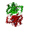
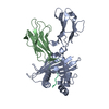
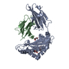
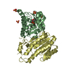
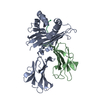
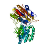
- PDB-4jp3: Crystal Structure of TT0495 protein from Thermus thermophilus HB8 -
+ Open data

- Basic information
| Entry | Database: PDB / ID: 4jp3 | ||||||
|---|---|---|---|---|---|---|---|
| Title | Crystal Structure of TT0495 protein from Thermus thermophilus HB8 | ||||||
 Components Components | 2-deoxy-D-gluconate 3-dehydrogenase | ||||||
 Keywords Keywords | OXIDOREDUCTASE / Rossmann fold | ||||||
| Function / homology |  Function and homology information Function and homology informationoxidoreductase activity, acting on the CH-OH group of donors, NAD or NADP as acceptor / nucleotide binding Similarity search - Function | ||||||
| Biological species |   Thermus thermophilus (bacteria) Thermus thermophilus (bacteria) | ||||||
| Method |  X-RAY DIFFRACTION / SYNCHROTRON / MOLECULAR REPLACEMENT / Resolution: 1.5 Å X-RAY DIFFRACTION / SYNCHROTRON / MOLECULAR REPLACEMENT / Resolution: 1.5 Å | ||||||
 Authors Authors | Pampa, K.J. / Lokanath, N.K. / Kunishima, N. / Ravishnkar Rai, V. | ||||||
 Citation Citation | Journal: Acta Crystallogr.,Sect.D / Year: 2014 Title: The first crystal structure of NAD-dependent 3-dehydro-2-deoxy-D-gluconate dehydrogenase from Thermus thermophilus HB8 Authors: Pampa, K.J. / Lokanath, N.K. / Kunishima, N. / Ravishnkar Rai, V. | ||||||
| History |
|
- Structure visualization
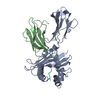
| Structure viewer | Molecule: MolmilJmol/JSmol |
|---|
- Downloads & links
-Download
| PDBx/mmCIF format | 4jp3.cif.gz | 64.8 KB | Display | PDBx/mmCIF format |
|---|---|---|---|---|
| PDB format | pdb4jp3.ent.gz | 47.1 KB | Display | PDB format |
| PDBx/mmJSON format | 4jp3.json.gz | Tree view | PDBx/mmJSON format | |
| Others |  Other downloads Other downloads |
-Validation report
| Arichive directory | https://data.pdbj.org/pub/pdb/validation_reports/jp/4jp3ftp://data.pdbj.org/pub/pdb/validation_reports/jp/4jp3 | HTTPS FTP |
|---|
-Related structure data
| Related structure data |  2ekpC  2ekqC 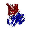 4jp2C 1x1eS C: citing same article ( S: Starting model for refinement |
|---|---|
| Similar structure data |


-Links
 PDBj
PDBj

- Assembly
| Deposited unit | 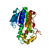
| ||||||||
|---|---|---|---|---|---|---|---|---|---|
| 1 | 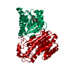
| ||||||||
| 2 | 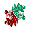
| ||||||||
| 3 | 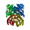
| ||||||||
| Unit cell |
|
-Components
| #1: Protein | Mass: 25702.729 Da / Num. of mol.: 1 / Source method: isolated from a natural source / Source: (natural) Thermus thermophilus (bacteria) / Strain: HB8 / References: UniProt: Q53W82 |
|---|---|
| #2: Chemical | ChemComp-CIT /   Mass: 192.124 Da / Num. of mol.: 1 / Source method: obtained synthetically / Formula: C6H8O7 Mass: 192.124 Da / Num. of mol.: 1 / Source method: obtained synthetically / Formula: C6H8O7 |
| #3: Water | ChemComp-HOH /  Mass: 18.015 Da / Num. of mol.: 324 / Source method: isolated from a natural source / Formula: H2O Mass: 18.015 Da / Num. of mol.: 324 / Source method: isolated from a natural source / Formula: H2O |
-Experimental details
-Experiment
| Experiment | Method: X-RAY DIFFRACTION / Number of used crystals: 1 |
|---|
- Sample preparation
| Crystal | Density Matthews: 2.59 Å3/Da / Density % sol: 52.59 % |
|---|---|
| Crystal grow | Temperature: 298 K / Method: evaporation / pH: 7.9 Details: 15% PEG 2000, 1.1M MgCl2, 0.1M NaCl, pH 7.9, EVAPORATION, temperature 298.0K |
-Data collection
| Diffraction | Mean temperature: 100 K |
|---|---|
| Diffraction source | Source: SYNCHROTRON / Site: SPring-8  / Beamline: BL26B2 / Wavelength: 0.9741 Å / Beamline: BL26B2 / Wavelength: 0.9741 Å |
| Detector | Type: RIGAKU / Detector: IMAGE PLATE / Date: Feb 18, 2013 |
| Radiation | Monochromator: graphite / Protocol: SINGLE WAVELENGTH / Monochromatic (M) / Laue (L): M / Scattering type: x-ray |
| Radiation wavelength | Wavelength: 0.9741 Å / Relative weight: 1 |
| Reflection | Resolution: 1.5→50 Å / Num. obs: 42860 / % possible obs: 99.4 % / Observed criterion σ(F): 1 / Observed criterion σ(I): 1 / Biso Wilson estimate: 13 Å2 |
| Reflection shell | Resolution: 1.5→1.55 Å / % possible all: 99.8 |
- Processing
| Software |
| |||||||||||||||||||||||||
|---|---|---|---|---|---|---|---|---|---|---|---|---|---|---|---|---|---|---|---|---|---|---|---|---|---|---|
| Refinement | Method to determine structure: MOLECULAR REPLACEMENT Starting model: 1x1e Resolution: 1.5→34.69 Å / Rfactor Rfree error: 0.004 / Data cutoff high absF: 1792496.25 / Data cutoff low absF: 0 / Isotropic thermal model: RESTRAINED / Cross valid method: THROUGHOUT / σ(F): 0
| |||||||||||||||||||||||||
| Solvent computation | Solvent model: FLAT MODEL / Bsol: 53.6891 Å2 / ksol: 0.4 e/Å3 | |||||||||||||||||||||||||
| Displacement parameters | Biso mean: 12.7 Å2
| |||||||||||||||||||||||||
| Refine analyze |
| |||||||||||||||||||||||||
| Refinement step | Cycle: LAST / Resolution: 1.5→34.69 Å
| |||||||||||||||||||||||||
| Refine LS restraints |
| |||||||||||||||||||||||||
| LS refinement shell | Resolution: 1.5→1.59 Å / Rfactor Rfree error: 0.013 / Total num. of bins used: 6
|
