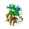
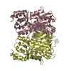
polyketide synthase complex / chalcone biosynthetic process / DIM/DIP cell wall layer assembly / acyltransferase activity, transferring groups other than amino-acyl groups / lipid biosynthetic process / Transferases; Acyltransferases; Transferring groups other than aminoacyl groups / fatty acid biosynthetic process Similarity search - Function
Chalcone/stilbene synthase, N-terminal / Chalcone and stilbene synthases, N-terminal domain / Polyketide synthase, type III / Chalcone/stilbene synthase, C-terminal / Chalcone and stilbene synthases, C-terminal domain / Thiolase/Chalcone synthase / Peroxisomal Thiolase; Chain A, domain 1 / Thiolase-like / 3-Layer(aba) Sandwich / Alpha Beta Similarity search - Domain/homology
Alpha-pyronesynthesispolyketidesynthase-likePks11 / Alpha-pyrone synthesis polyketide synthase type III Pks11 / Chalcone synthase-like protein / CHS-like
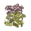
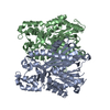
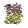
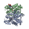
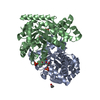
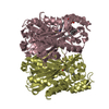
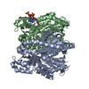
Mass: 37681.145 Da / Num. of mol.: 4 Source method: isolated from a genetically manipulated source Source: (gene. exp.) Mycobacterium tuberculosis (bacteria) / Strain: H37RV / Gene: pks11, Rv1665, MT1705 / Production host: mycobacterium smegmatis (bacteria) References: UniProt: O06587, UniProt: P9WPF3*PLUS, Transferases; Acyltransferases; Transferring groups other than aminoacyl groups
In the structure databanks used in Yorodumi, some data are registered as the other names, "COVID-19 virus" and "2019-nCoV". Here are the details of the virus and the list of structure data.
Jan 31, 2019. EMDB accession codes are about to change! (news from PDBe EMDB page)
EMDB accession codes are about to change! (news from PDBe EMDB page)
The allocation of 4 digits for EMDB accession codes will soon come to an end. Whilst these codes will remain in use, new EMDB accession codes will include an additional digit and will expand incrementally as the available range of codes is exhausted. The current 4-digit format prefixed with “EMD-” (i.e. EMD-XXXX) will advance to a 5-digit format (i.e. EMD-XXXXX), and so on. It is currently estimated that the 4-digit codes will be depleted around Spring 2019, at which point the 5-digit format will come into force.
The EM Navigator/Yorodumi systems omit the EMD- prefix.
Related info.:Q: What is EMD? / ID/Accession-code notation in Yorodumi/EM Navigator
Yorodumi is a browser for structure data from EMDB, PDB, SASBDB, etc.
This page is also the successor to EM Navigator detail page, and also detail information page/front-end page for Omokage search.
The word "yorodu" (or yorozu) is an old Japanese word meaning "ten thousand". "mi" (miru) is to see.
Related info.:EMDB / PDB / SASBDB / Comparison of 3 databanks / Yorodumi Search / Aug 31, 2016. New EM Navigator & Yorodumi / Yorodumi Papers / Jmol/JSmol / Function and homology information / Changes in new EM Navigator and Yorodumi
 Movie
Movie Controller

 Yorodumi
 Open data
 Basic information
 Components
 Keywords
 Function and homology information
 Mycobacterium tuberculosis (bacteria)
 X-RAY DIFFRACTION /
  Authors
 Citation
 Structure visualization
 Downloads & links
 Other downloads


 PDBj
PDBj


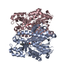
 Assembly
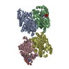

 Mass: 256.424 Da / Num. of mol.: 2 / Source method: obtained synthetically / Formula: C16H32O2
 Mass: 767.534 Da / Num. of mol.: 2 / Source method: obtained synthetically / Formula: C21H36N7O16P3S
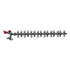
 Mass: 312.487 Da / Num. of mol.: 2 / Source method: obtained synthetically / Formula: C19H36O3
 Mass: 18.015 Da / Num. of mol.: 185 / Source method: isolated from a natural source / Formula: H2O
 Sample preparation
 / Beamline: 23-ID-B / Wavelength: 1 Å
 Processing