 Movie
Movie Controller

[English] 日本語
 Yorodumi
- PDB-4j3s: Crystal structure of barley limit dextrinase soaked with 300mM ma... -
+ Open data

- Basic information
| Entry | Database: PDB / ID: 4j3s | |||||||||
|---|---|---|---|---|---|---|---|---|---|---|
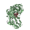
| Title | Crystal structure of barley limit dextrinase soaked with 300mM maltotetraose | |||||||||
 Components Components | Limit dextrinase | |||||||||
 Keywords Keywords | HYDROLASE / GH13 hydrolase | |||||||||
| Function / homology |  Function and homology information Function and homology informationpullulanase activity / polysaccharide catabolic process / metal ion binding Similarity search - Function | |||||||||
| Biological species |  | |||||||||
| Method |  X-RAY DIFFRACTION / SYNCHROTRON / MOLECULAR REPLACEMENT / Resolution: 1.75 Å X-RAY DIFFRACTION / SYNCHROTRON / MOLECULAR REPLACEMENT / Resolution: 1.75 Å | |||||||||
 Authors Authors | Sim, L. / Windahl, M.S. / Moeller, M.S. / Henriksen, A. | |||||||||
 Citation Citation | Journal: J.Mol.Biol. / Year: 2015 Title: Oligosaccharide and substrate binding in the starch debranching enzyme barley limit dextrinase Authors: Moeller, M.S. / Windahl, M.S. / Sim, L. / Bjstrup, M. / Abou Hachem, M. / Hindsgaul, O. / Palcic, M. / Svensson, B. / Henriksen, A. | |||||||||
| History |
|
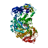
- Structure visualization
| Structure viewer | Molecule: MolmilJmol/JSmol |
|---|
- Downloads & links
-Download
| PDBx/mmCIF format | 4j3s.cif.gz | 370 KB | Display | PDBx/mmCIF format |
|---|---|---|---|---|
| PDB format | pdb4j3s.ent.gz | 296.2 KB | Display | PDB format |
| PDBx/mmJSON format | 4j3s.json.gz | Tree view | PDBx/mmJSON format | |
| Others |  Other downloads Other downloads |
-Validation report
| Arichive directory | https://data.pdbj.org/pub/pdb/validation_reports/j3/4j3sftp://data.pdbj.org/pub/pdb/validation_reports/j3/4j3s | HTTPS FTP |
|---|
-Related structure data
| Related structure data |  4j3tC 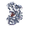 4j3uC  4j3vC  4j3wC 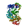 4j3xC  2y4sS C: citing same article ( S: Starting model for refinement |
|---|---|
| Similar structure data |

-Links
 PDBj
PDBj- Assembly
| Deposited unit | 
| ||||||||||||
|---|---|---|---|---|---|---|---|---|---|---|---|---|---|
| 1 |
| ||||||||||||
| Unit cell |
| ||||||||||||
| Components on special symmetry positions |
|
-Components
-Protein , 1 types, 1 molecules A
| #1: Protein | Mass: 99746.727 Da / Num. of mol.: 1 / Fragment: UNP Residues 22-905 Source method: isolated from a genetically manipulated source Source: (gene. exp.)  |
|---|
-Sugars , 2 types, 3 molecules
| #2: Polysaccharide | 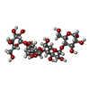  Source method: isolated from a genetically manipulated source Details: oligosaccharide / References: alpha-maltotetraose #3: Polysaccharide | alpha-D-glucopyranose-(1-4)-alpha-D-glucopyranose-(1-4)-alpha-D-glucopyranose / alpha-maltotriose |   Source method: isolated from a genetically manipulated source Details: oligosaccharide / References: alpha-maltotriose |
|---|
-Non-polymers , 4 types, 546 molecules 


| #4: Chemical | ChemComp-IOD /  Mass: 126.904 Da / Num. of mol.: 6 / Source method: obtained synthetically / Formula: I Mass: 126.904 Da / Num. of mol.: 6 / Source method: obtained synthetically / Formula: I#5: Chemical | ChemComp-CA / |  Mass: 40.078 Da / Num. of mol.: 1 / Source method: obtained synthetically / Formula: Ca Mass: 40.078 Da / Num. of mol.: 1 / Source method: obtained synthetically / Formula: Ca#6: Chemical | ChemComp-GOL / |  Mass: 92.094 Da / Num. of mol.: 1 / Source method: obtained synthetically / Formula: C3H8O3 Mass: 92.094 Da / Num. of mol.: 1 / Source method: obtained synthetically / Formula: C3H8O3#7: Water | ChemComp-HOH / | Mass: 18.015 Da / Num. of mol.: 538 / Source method: isolated from a natural source / Formula: H2O |
|---|
-Details
| Sequence details | THESE RESIDUES REPRESENT NATURAL VARIANTS. |
|---|
-Experimental details
-Experiment
| Experiment | Method: X-RAY DIFFRACTION / Number of used crystals: 1 |
|---|
- Sample preparation
| Crystal | Density Matthews: 1.97 Å3/Da / Density % sol: 37.53 % |
|---|---|
| Crystal grow | Temperature: 298 K / Method: vapor diffusion, hanging drop Details: 20% PEG 3350, 0.3M NaI, VAPOR DIFFUSION, HANGING DROP, temperature 298K |
-Data collection
| Diffraction | Mean temperature: 100 K | ||||||||||||||||||||||||||||||||||||||||||||||||||||||||||||||||||||||||||||||||||||||||||||||||||||
|---|---|---|---|---|---|---|---|---|---|---|---|---|---|---|---|---|---|---|---|---|---|---|---|---|---|---|---|---|---|---|---|---|---|---|---|---|---|---|---|---|---|---|---|---|---|---|---|---|---|---|---|---|---|---|---|---|---|---|---|---|---|---|---|---|---|---|---|---|---|---|---|---|---|---|---|---|---|---|---|---|---|---|---|---|---|---|---|---|---|---|---|---|---|---|---|---|---|---|---|---|---|
| Diffraction source | Source: SYNCHROTRON / Site: ESRF  / Beamline: ID23-1 / Wavelength: 0.992 Å / Beamline: ID23-1 / Wavelength: 0.992 Å | ||||||||||||||||||||||||||||||||||||||||||||||||||||||||||||||||||||||||||||||||||||||||||||||||||||
| Detector | Type: ADSC QUANTUM 315r / Detector: CCD / Date: Jun 1, 2012 | ||||||||||||||||||||||||||||||||||||||||||||||||||||||||||||||||||||||||||||||||||||||||||||||||||||
| Radiation | Protocol: SINGLE WAVELENGTH / Monochromatic (M) / Laue (L): M / Scattering type: x-ray | ||||||||||||||||||||||||||||||||||||||||||||||||||||||||||||||||||||||||||||||||||||||||||||||||||||
| Radiation wavelength | Wavelength: 0.992 Å / Relative weight: 1 | ||||||||||||||||||||||||||||||||||||||||||||||||||||||||||||||||||||||||||||||||||||||||||||||||||||
| Reflection | Resolution: 1.75→50 Å / Num. obs: 77622 / % possible obs: 98.8 % / Observed criterion σ(I): -3 / Biso Wilson estimate: 27.005 Å2 / Rmerge(I) obs: 0.06 / Net I/σ(I): 14.44 | ||||||||||||||||||||||||||||||||||||||||||||||||||||||||||||||||||||||||||||||||||||||||||||||||||||
| Reflection shell | Diffraction-ID: 1
|
- Processing
| Software |
| |||||||||||||||||||||||||||||||||||||||||||||||||||||||||||||||||||||||||||
|---|---|---|---|---|---|---|---|---|---|---|---|---|---|---|---|---|---|---|---|---|---|---|---|---|---|---|---|---|---|---|---|---|---|---|---|---|---|---|---|---|---|---|---|---|---|---|---|---|---|---|---|---|---|---|---|---|---|---|---|---|---|---|---|---|---|---|---|---|---|---|---|---|---|---|---|---|
| Refinement | Method to determine structure: MOLECULAR REPLACEMENT Starting model: 2Y4S Resolution: 1.75→46.96 Å / Cor.coef. Fo:Fc: 0.972 / Cor.coef. Fo:Fc free: 0.955 / WRfactor Rfree: 0.1778 / WRfactor Rwork: 0.1344 / Occupancy max: 1 / Occupancy min: 0.5 / FOM work R set: 0.9031 / SU B: 4.665 / SU ML: 0.067 / SU R Cruickshank DPI: 0.2415 / SU Rfree: 0.1025 / Cross valid method: THROUGHOUT / σ(F): 0 / ESU R: 0.241 / ESU R Free: 0.102 / Stereochemistry target values: MAXIMUM LIKELIHOOD Details: HYDROGENS HAVE BEEN ADDED IN THE RIDING POSITIONS U VALUES: REFINED INDIVIDUALLY
| |||||||||||||||||||||||||||||||||||||||||||||||||||||||||||||||||||||||||||
| Solvent computation | Ion probe radii: 0.8 Å / Shrinkage radii: 0.8 Å / VDW probe radii: 1.2 Å / Solvent model: MASK | |||||||||||||||||||||||||||||||||||||||||||||||||||||||||||||||||||||||||||
| Displacement parameters | Biso max: 122.12 Å2 / Biso mean: 23.1793 Å2 / Biso min: 9.03 Å2
| |||||||||||||||||||||||||||||||||||||||||||||||||||||||||||||||||||||||||||
| Refinement step | Cycle: LAST / Resolution: 1.75→46.96 Å
| |||||||||||||||||||||||||||||||||||||||||||||||||||||||||||||||||||||||||||
| Refine LS restraints |
| |||||||||||||||||||||||||||||||||||||||||||||||||||||||||||||||||||||||||||
| LS refinement shell | Resolution: 1.75→1.795 Å / Total num. of bins used: 20
|
