 Movie
Movie Controller


+ Open data

- Basic information
| Entry | Database: PDB / ID: 4iyl | |||||||||
|---|---|---|---|---|---|---|---|---|---|---|
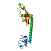
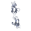
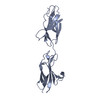
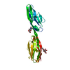
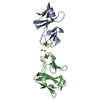
| Title | 30S ribosomal protein S15 from Campylobacter jejuni | |||||||||
 Components Components | 30S ribosomal protein S15 | |||||||||
 Keywords Keywords | RIBOSOMAL PROTEIN / structural genomics / Center for Structural Genomics of Infectious Diseases / CSGID / rRNA binding / translation / NIAID / National Institute of Allergy and Infectious Diseases | |||||||||
| Function / homology |  Function and homology information Function and homology informationcytosolic small ribosomal subunit / rRNA binding / structural constituent of ribosome / translation Similarity search - Function | |||||||||
| Biological species |  Campylobacter jejuni subsp. jejuni (Campylobacter) Campylobacter jejuni subsp. jejuni (Campylobacter) | |||||||||
| Method |  X-RAY DIFFRACTION / SYNCHROTRON / MOLECULAR REPLACEMENT / Resolution: 2.36 Å X-RAY DIFFRACTION / SYNCHROTRON / MOLECULAR REPLACEMENT / Resolution: 2.36 Å | |||||||||
 Authors Authors | Osipiuk, J. / Nocek, B. / Gu, M. / Kwon, K. / Anderson, W.F. / Joachimiak, A. / Center for Structural Genomics of Infectious Diseases (CSGID) | |||||||||
 Citation Citation | Journal: To be Published Title: 30S ribosomal protein S15 from Campylobacter jejuni Authors: Osipiuk, J. / Nocek, B. / Gu, M. / Kwon, K. / Anderson, W.F. / Joachimiak, A. | |||||||||
| History |
|
- Structure visualization
| Structure viewer | Molecule: MolmilJmol/JSmol |
|---|
- Downloads & links
-Download
| PDBx/mmCIF format | 4iyl.cif.gz | 29.2 KB | Display | PDBx/mmCIF format |
|---|---|---|---|---|
| PDB format | pdb4iyl.ent.gz | 18.7 KB | Display | PDB format |
| PDBx/mmJSON format | 4iyl.json.gz | Tree view | PDBx/mmJSON format | |
| Others |  Other downloads Other downloads |
-Validation report
| Summary document | 4iyl_validation.pdf.gz | 429.9 KB | Display | wwPDB validaton report |
|---|---|---|---|---|
| Full document | 4iyl_full_validation.pdf.gz | 430.3 KB | Display | |
| Data in XML | 4iyl_validation.xml.gz | 5.2 KB | Display | |
| Data in CIF | 4iyl_validation.cif.gz | 6.2 KB | Display | |
| Arichive directory | https://data.pdbj.org/pub/pdb/validation_reports/iy/4iylftp://data.pdbj.org/pub/pdb/validation_reports/iy/4iyl | HTTPS FTP |
-Related structure data
| Related structure data |  2y0y S: Starting model for refinement |
|---|---|
| Similar structure data | |
| Other databases |

-Links
 PDBj
PDBj- Assembly
| Deposited unit | 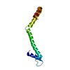
| ||||||||
|---|---|---|---|---|---|---|---|---|---|
| 1 |
| ||||||||
| 2 | 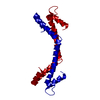
| ||||||||
| 3 | 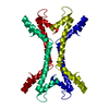
| ||||||||
| Unit cell |
| ||||||||
| Details | biological unit is the same as asymmetric unit, based on ribosome structure |
-Components
| #1: Protein | Mass: 10519.337 Da / Num. of mol.: 1 Source method: isolated from a genetically manipulated source Source: (gene. exp.) Campylobacter jejuni subsp. jejuni (Campylobacter)Strain: NCTC 11168 / Gene: Cj0884, rpsO / Plasmid: pMCSG7 / Production host: |
|---|---|
| #2: Water | ChemComp-HOH /  Mass: 18.015 Da / Num. of mol.: 4 / Source method: isolated from a natural source / Formula: H2O Mass: 18.015 Da / Num. of mol.: 4 / Source method: isolated from a natural source / Formula: H2O |
-Experimental details
-Experiment
| Experiment | Method: X-RAY DIFFRACTION / Number of used crystals: 1 |
|---|
- Sample preparation
| Crystal | Density Matthews: 3.49 Å3/Da / Density % sol: 64.76 % |
|---|---|
| Crystal grow | Temperature: 289 K / Method: vapor diffusion, sitting drop / pH: 7.1 Details: 0.49 M sodium phosphate, 0.91 M di-potassium phosphate, pH 7.1, VAPOR DIFFUSION, SITTING DROP, temperature 289K |
-Data collection
| Diffraction | Mean temperature: 100 K | |||||||||||||||||||||||||||||||||||||||||||||||||||||||||||||||||||||||||||||||||||||||||||||||||||||||||||||||||||||||||||||||||||||||||||||||||||
|---|---|---|---|---|---|---|---|---|---|---|---|---|---|---|---|---|---|---|---|---|---|---|---|---|---|---|---|---|---|---|---|---|---|---|---|---|---|---|---|---|---|---|---|---|---|---|---|---|---|---|---|---|---|---|---|---|---|---|---|---|---|---|---|---|---|---|---|---|---|---|---|---|---|---|---|---|---|---|---|---|---|---|---|---|---|---|---|---|---|---|---|---|---|---|---|---|---|---|---|---|---|---|---|---|---|---|---|---|---|---|---|---|---|---|---|---|---|---|---|---|---|---|---|---|---|---|---|---|---|---|---|---|---|---|---|---|---|---|---|---|---|---|---|---|---|---|---|---|
| Diffraction source | Source: SYNCHROTRON / Site: APS  / Beamline: 19-ID / Wavelength: 0.9792 Å / Beamline: 19-ID / Wavelength: 0.9792 Å | |||||||||||||||||||||||||||||||||||||||||||||||||||||||||||||||||||||||||||||||||||||||||||||||||||||||||||||||||||||||||||||||||||||||||||||||||||
| Detector | Type: ADSC QUANTUM 315r / Detector: CCD / Date: Aug 25, 2010 | |||||||||||||||||||||||||||||||||||||||||||||||||||||||||||||||||||||||||||||||||||||||||||||||||||||||||||||||||||||||||||||||||||||||||||||||||||
| Radiation | Monochromator: double crystal monochromator / Protocol: SINGLE WAVELENGTH / Monochromatic (M) / Laue (L): M / Scattering type: x-ray | |||||||||||||||||||||||||||||||||||||||||||||||||||||||||||||||||||||||||||||||||||||||||||||||||||||||||||||||||||||||||||||||||||||||||||||||||||
| Radiation wavelength | Wavelength: 0.9792 Å / Relative weight: 1 | |||||||||||||||||||||||||||||||||||||||||||||||||||||||||||||||||||||||||||||||||||||||||||||||||||||||||||||||||||||||||||||||||||||||||||||||||||
| Reflection | Resolution: 2.36→32.3 Å / Num. all: 5992 / Num. obs: 5992 / % possible obs: 95.2 % / Observed criterion σ(F): 0 / Observed criterion σ(I): 0 / Redundancy: 3.6 % / Biso Wilson estimate: 68.7 Å2 / Rmerge(I) obs: 0.098 / Χ2: 1.39 / Net I/σ(I): 10.2 | |||||||||||||||||||||||||||||||||||||||||||||||||||||||||||||||||||||||||||||||||||||||||||||||||||||||||||||||||||||||||||||||||||||||||||||||||||
| Reflection shell | Diffraction-ID: 1
|
- Processing
| Software |
| ||||||||||||||||||||||||||||||||||||||||||||||||||||||||||||
|---|---|---|---|---|---|---|---|---|---|---|---|---|---|---|---|---|---|---|---|---|---|---|---|---|---|---|---|---|---|---|---|---|---|---|---|---|---|---|---|---|---|---|---|---|---|---|---|---|---|---|---|---|---|---|---|---|---|---|---|---|---|
| Refinement | Method to determine structure: MOLECULAR REPLACEMENT Starting model: PDB ENTRY 2Y0Y 2y0y Resolution: 2.36→32.27 Å / Cor.coef. Fo:Fc: 0.942 / Cor.coef. Fo:Fc free: 0.929 / Occupancy max: 1 / Occupancy min: 1 / SU B: 9.575 / SU ML: 0.197 / Cross valid method: THROUGHOUT / σ(F): 0 / σ(I): 0 / ESU R: 0.287 / ESU R Free: 0.237 / Stereochemistry target values: MAXIMUM LIKELIHOOD Details: HYDROGENS HAVE BEEN ADDED IN THE RIDING POSITIONS U VALUES : REFINED INDIVIDUALLY
| ||||||||||||||||||||||||||||||||||||||||||||||||||||||||||||
| Solvent computation | Ion probe radii: 0.8 Å / Shrinkage radii: 0.8 Å / VDW probe radii: 1.2 Å / Solvent model: MASK | ||||||||||||||||||||||||||||||||||||||||||||||||||||||||||||
| Displacement parameters | Biso max: 151.27 Å2 / Biso mean: 81.4788 Å2 / Biso min: 45.6 Å2
| ||||||||||||||||||||||||||||||||||||||||||||||||||||||||||||
| Refinement step | Cycle: LAST / Resolution: 2.36→32.27 Å
| ||||||||||||||||||||||||||||||||||||||||||||||||||||||||||||
| Refine LS restraints |
| ||||||||||||||||||||||||||||||||||||||||||||||||||||||||||||
| LS refinement shell | Resolution: 2.362→2.423 Å / Total num. of bins used: 20
|
