 Movie
Movie Controller


+ Open data

- Basic information
| Entry | Database: PDB / ID: 4ipg | ||||||
|---|---|---|---|---|---|---|---|
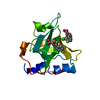
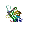
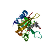
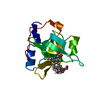
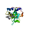
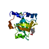
| Title | Structure of the N-terminal domain of RPA70, E7R, E100R mutant | ||||||
 Components Components | Replication protein A 70 kDa DNA-binding subunit | ||||||
 Keywords Keywords | PROTEIN BINDING / OB-fold | ||||||
| Function / homology |  Function and homology information Function and homology informationprotein localization to chromosome / DNA replication factor A complex / single-stranded telomeric DNA binding / Removal of the Flap Intermediate / lateral element / G-rich strand telomeric DNA binding / protein localization to site of double-strand break / Mismatch repair (MMR) directed by MSH2:MSH3 (MutSbeta) / Mismatch repair (MMR) directed by MSH2:MSH6 (MutSalpha) / chromatin-protein adaptor activity ...protein localization to chromosome / DNA replication factor A complex / single-stranded telomeric DNA binding / Removal of the Flap Intermediate / lateral element / G-rich strand telomeric DNA binding / protein localization to site of double-strand break / Mismatch repair (MMR) directed by MSH2:MSH3 (MutSbeta) / Mismatch repair (MMR) directed by MSH2:MSH6 (MutSalpha) / chromatin-protein adaptor activity / Removal of the Flap Intermediate from the C-strand / HDR through Single Strand Annealing (SSA) / Impaired BRCA2 binding to RAD51 / hemopoiesis / PCNA-Dependent Long Patch Base Excision Repair / Activation of the pre-replicative complex / Regulation of HSF1-mediated heat shock response / Presynaptic phase of homologous DNA pairing and strand exchange / HSF1 activation / Activation of ATR in response to replication stress / telomere maintenance via telomerase / mismatch repair / SUMOylation of DNA damage response and repair proteins / homeostasis of number of cells within a tissue / site of DNA damage / telomere maintenance / male germ cell nucleus / Translesion synthesis by REV1 / Translesion synthesis by POLK / Translesion synthesis by POLI / Gap-filling DNA repair synthesis and ligation in GG-NER / nucleotide-excision repair / Fanconi Anemia Pathway / Termination of translesion DNA synthesis / meiotic cell cycle / Translesion Synthesis by POLH / base-excision repair / PML body / Recognition of DNA damage by PCNA-containing replication complex / G2/M DNA damage checkpoint / double-strand break repair via homologous recombination / DNA-templated DNA replication / Meiotic recombination / HDR through Homologous Recombination (HRR) / Dual Incision in GG-NER / Formation of Incision Complex in GG-NER / Dual incision in TC-NER / Gap-filling DNA repair synthesis and ligation in TC-NER / single-stranded DNA binding / site of double-strand break / in utero embryonic development / Processing of DNA double-strand break ends / DNA recombination / Regulation of TP53 Activity through Phosphorylation / damaged DNA binding / chromosome, telomeric region / DNA replication / DNA repair / chromatin binding / positive regulation of cell population proliferation / DNA damage response / zinc ion binding / nucleoplasm / nucleus Similarity search - Function | ||||||
| Biological species |  Homo sapiens (human) Homo sapiens (human) | ||||||
| Method |  X-RAY DIFFRACTION / SYNCHROTRON / MOLECULAR REPLACEMENT / Resolution: 1.58 Å X-RAY DIFFRACTION / SYNCHROTRON / MOLECULAR REPLACEMENT / Resolution: 1.58 Å | ||||||
 Authors Authors | Feldkamp, M.D. / Frank, A.O. / Vangamudi, B. / Fesik, S.W. / Chazin, W.J. | ||||||
 Citation Citation | Journal: Biochemistry / Year: 2013 Title: Surface Reengineering of RPA70N Enables Cocrystallization with an Inhibitor of the Replication Protein A Interaction Motif of ATR Interacting Protein. Authors: Feldkamp, M.D. / Frank, A.O. / Kennedy, J.P. / Patrone, J.D. / Vangamudi, B. / Waterson, A.G. / Fesik, S.W. / Chazin, W.J. | ||||||
| History |
|
- Structure visualization
| Structure viewer | Molecule: MolmilJmol/JSmol |
|---|
- Downloads & links
-Download
| PDBx/mmCIF format | 4ipg.cif.gz | 63.3 KB | Display | PDBx/mmCIF format |
|---|---|---|---|---|
| PDB format | pdb4ipg.ent.gz | 47.4 KB | Display | PDB format |
| PDBx/mmJSON format | 4ipg.json.gz | Tree view | PDBx/mmJSON format | |
| Others |  Other downloads Other downloads |
-Validation report
| Arichive directory | https://data.pdbj.org/pub/pdb/validation_reports/ip/4ipgftp://data.pdbj.org/pub/pdb/validation_reports/ip/4ipg | HTTPS FTP |
|---|
-Related structure data
| Related structure data | 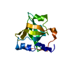 4ipcC 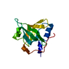 4ipdC 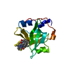 4iphC 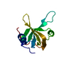 2b29S C: citing same article ( S: Starting model for refinement |
|---|---|
| Similar structure data |


-Links
 PDBj
PDBj


- Assembly
| Deposited unit | 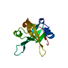
| ||||||||
|---|---|---|---|---|---|---|---|---|---|
| 1 |
| ||||||||
| Unit cell |
|
-Components
| #1: Protein | Mass: 13525.808 Da / Num. of mol.: 1 / Mutation: E7R, E100R Source method: isolated from a genetically manipulated source Source: (gene. exp.) Homo sapiens (human) / Gene: RPA1, REPA1, RPA70 / Plasmid: pET15b / Production host:  |
|---|---|
| #2: Water | ChemComp-HOH /  Mass: 18.015 Da / Num. of mol.: 148 / Source method: isolated from a natural source / Formula: H2O Mass: 18.015 Da / Num. of mol.: 148 / Source method: isolated from a natural source / Formula: H2O |
-Experimental details
-Experiment
| Experiment | Method: X-RAY DIFFRACTION / Number of used crystals: 1 |
|---|
- Sample preparation
| Crystal | Density Matthews: 2.08 Å3/Da / Density % sol: 40.89 % |
|---|---|
| Crystal grow | Temperature: 294 K / Method: vapor diffusion, hanging drop / pH: 5 Details: 100 mM Citrate, 200 mM NaCl, 30% PEG 8000, pH 5.0, VAPOR DIFFUSION, HANGING DROP, temperature 294K |
-Data collection
| Diffraction | Mean temperature: 100 K |
|---|---|
| Diffraction source | Source: SYNCHROTRON / Site: APS  / Beamline: 21-ID-D / Wavelength: 0.97857 Å / Beamline: 21-ID-D / Wavelength: 0.97857 Å |
| Detector | Type: MAR scanner 300 mm plate / Detector: IMAGE PLATE / Date: May 22, 2012 |
| Radiation | Monochromator: Si(111) / Protocol: SINGLE WAVELENGTH / Monochromatic (M) / Laue (L): M / Scattering type: x-ray |
| Radiation wavelength | Wavelength: 0.97857 Å / Relative weight: 1 |
| Reflection | Resolution: 1.58→24.266 Å / Num. obs: 16007 / % possible obs: 99.92 % / Observed criterion σ(F): 1.58 / Observed criterion σ(I): 1.58 / Redundancy: 5.9 % / Biso Wilson estimate: 19.8 Å2 / Rsym value: 0.065 / Net I/σ(I): 18.3 |
| Reflection shell | Resolution: 1.58→1.6794 Å / % possible all: 100 |
- Processing
| Software | Name: PHENIX / Version: (phenix.refine: 1.8.1_1168) / Classification: refinement | |||||||||||||||||||||||||||||||||||||||||||||||||
|---|---|---|---|---|---|---|---|---|---|---|---|---|---|---|---|---|---|---|---|---|---|---|---|---|---|---|---|---|---|---|---|---|---|---|---|---|---|---|---|---|---|---|---|---|---|---|---|---|---|---|
| Refinement | Method to determine structure: MOLECULAR REPLACEMENT Starting model: 2B29 Resolution: 1.58→24.266 Å / SU ML: 0.14 / σ(F): 1.34 / Phase error: 19.11 / Stereochemistry target values: ML
| |||||||||||||||||||||||||||||||||||||||||||||||||
| Solvent computation | Shrinkage radii: 0.8 Å / VDW probe radii: 1 Å / Solvent model: FLAT BULK SOLVENT MODEL | |||||||||||||||||||||||||||||||||||||||||||||||||
| Refinement step | Cycle: LAST / Resolution: 1.58→24.266 Å
| |||||||||||||||||||||||||||||||||||||||||||||||||
| Refine LS restraints |
| |||||||||||||||||||||||||||||||||||||||||||||||||
| LS refinement shell |
|
