 Movie
Movie Controller

[English] 日本語
 Yorodumi
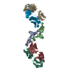
- PDB-4ild: Crystal structure of truncated Bovine viral diarrhea virus 1 E2 e... -
+ Open data

- Basic information
| Entry | Database: PDB / ID: 4ild | |||||||||
|---|---|---|---|---|---|---|---|---|---|---|
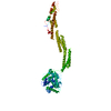
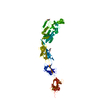
| Title | Crystal structure of truncated Bovine viral diarrhea virus 1 E2 envelope protein | |||||||||
 Components Components | Envelope glycoprotein E2 | |||||||||
 Keywords Keywords | VIRAL PROTEIN / BVDV1 / viral envelope protein / viral membrane fusion / E1 envelope protein / viral surface membrane | |||||||||
| Function / homology |  Function and homology information Function and homology informationpestivirus NS3 polyprotein peptidase / dopamine receptor binding / ribonuclease T2 / serine-type exopeptidase activity / DNA/DNA annealing activity / ribonuclease T2 activity / RNA strand annealing activity / viral genome packaging / RNA stabilization / host cell cytoplasmic vesicle ...pestivirus NS3 polyprotein peptidase / dopamine receptor binding / ribonuclease T2 / serine-type exopeptidase activity / DNA/DNA annealing activity / ribonuclease T2 activity / RNA strand annealing activity / viral genome packaging / RNA stabilization / host cell cytoplasmic vesicle / protein-DNA complex / nucleoside-triphosphate phosphatase / channel activity / monoatomic ion transmembrane transport / clathrin-dependent endocytosis of virus by host cell / symbiont-mediated suppression of host cytoplasmic pattern recognition receptor signaling pathway via inhibition of IRF3 activity / Hydrolases; Acting on peptide bonds (peptidases); Cysteine endopeptidases / RNA helicase activity / viral protein processing / host cell endoplasmic reticulum membrane / RNA helicase / ribonucleoprotein complex / serine-type endopeptidase activity / symbiont-mediated activation of host autophagy / RNA-directed RNA polymerase / cysteine-type endopeptidase activity / viral RNA genome replication / RNA-directed RNA polymerase activity / fusion of virus membrane with host endosome membrane / virion attachment to host cell / GTP binding / virion membrane / ATP hydrolysis activity / proteolysis / DNA binding / RNA binding / ATP binding / identical protein binding Similarity search - Function | |||||||||
| Biological species |  Bovine viral diarrhea virus 1-NADL Bovine viral diarrhea virus 1-NADL | |||||||||
| Method |  X-RAY DIFFRACTION / SYNCHROTRON / INITIAL SAD PLUS MULTIPLE HA SITE REFINEMENT / Resolution: 3.27 Å X-RAY DIFFRACTION / SYNCHROTRON / INITIAL SAD PLUS MULTIPLE HA SITE REFINEMENT / Resolution: 3.27 Å | |||||||||
 Authors Authors | Modis, Y. / Li, Y. / Wang, J. | |||||||||
 Citation Citation | Journal: Proc.Natl.Acad.Sci.USA / Year: 2013 Title: Crystal structure of glycoprotein E2 from bovine viral diarrhea virus. Authors: Li, Y. / Wang, J. / Kanai, R. / Modis, Y. | |||||||||
| History |
|
- Structure visualization
| Structure viewer | Molecule: MolmilJmol/JSmol |
|---|
- Downloads & links
-Download
| PDBx/mmCIF format | 4ild.cif.gz | 213.9 KB | Display | PDBx/mmCIF format |
|---|---|---|---|---|
| PDB format | pdb4ild.ent.gz | 173.2 KB | Display | PDB format |
| PDBx/mmJSON format | 4ild.json.gz | Tree view | PDBx/mmJSON format | |
| Others |  Other downloads Other downloads |
-Validation report
| Arichive directory | https://data.pdbj.org/pub/pdb/validation_reports/il/4ildftp://data.pdbj.org/pub/pdb/validation_reports/il/4ild | HTTPS FTP |
|---|
-Related structure data


-Links
 PDBj
PDBj

- Assembly
| Deposited unit | 
| ||||||||
|---|---|---|---|---|---|---|---|---|---|
| 1 |
| ||||||||
| Unit cell |
|
-Components
| #1: Protein | Mass: 28716.760 Da / Num. of mol.: 2 Fragment: N-terminal truncated BVDV1 E2 envelope protein, UNP residues 781-1030 Source method: isolated from a genetically manipulated source Details: N-terminal His-tagged / Source: (gene. exp.) Bovine viral diarrhea virus 1-NADL / Plasmid: pAcgp67a / Production host:  Trichoplusia ni (cabbage looper) / References: UniProt: P19711 Trichoplusia ni (cabbage looper) / References: UniProt: P19711#2: Polysaccharide | 2-acetamido-2-deoxy-beta-D-glucopyranose-(1-4)-2-acetamido-2-deoxy-beta-D-glucopyranose Source method: isolated from a genetically manipulated source #3: Chemical | ChemComp-CA /   Mass: 40.078 Da / Num. of mol.: 4 / Source method: obtained synthetically / Formula: Ca Mass: 40.078 Da / Num. of mol.: 4 / Source method: obtained synthetically / Formula: Ca#4: Chemical | ChemComp-IUM /   Mass: 270.028 Da / Num. of mol.: 4 / Source method: obtained synthetically / Formula: O2U Mass: 270.028 Da / Num. of mol.: 4 / Source method: obtained synthetically / Formula: O2UHas protein modification | Y | |
|---|
-Experimental details
-Experiment
| Experiment | Method: X-RAY DIFFRACTION / Number of used crystals: 1 |
|---|
- Sample preparation
| Crystal | Density Matthews: 3.11 Å3/Da / Density % sol: 60.4 % |
|---|---|
| Crystal grow | Temperature: 294 K / pH: 5.5 Details: 10% polyethylene glycol 3350, 0.1 M Bis-tris pH 5.5, 0.05 M cesium chloride, 0.04 M calcium acetate, 10% glycerol., VAPOR DIFFUSION, HANGING DROP, temperature 294K |
-Data collection
| Diffraction | Mean temperature: 195 K |
|---|---|
| Diffraction source | Source: SYNCHROTRON / Site: NSLS  / Beamline: X25 / Wavelength: 1.2142 / Beamline: X25 / Wavelength: 1.2142 |
| Detector | Type: S/N 60-0107 / Detector: PILATUS 6M / Date: Oct 3, 2012 |
| Radiation | Protocol: SINGLE WAVELENGTH / Monochromatic (M) / Laue (L): M / Scattering type: x-ray |
| Radiation wavelength | Wavelength: 1.2142 Å / Relative weight: 1 |
| Reflection | Resolution: 3.27→50 Å / Num. obs: 11169 / % possible obs: 98.4 % / Observed criterion σ(I): 1.9 / Rsym value: 0.086 / Net I/σ(I): 12.08 |
| Reflection shell | Resolution: 3.27→3.36 Å / Redundancy: 1.72 % / Mean I/σ(I) obs: 69.4 / % possible all: 89.1 |
- Processing
| Software |
| |||||||||||||||||||||||||||||||||||||||||||||||||||||||||||||||||||||||||||||||||||||||||||||||||||||||||||||||||||||||||||||||||||||||||||||||||||||||||||||||||||||||||||||||
|---|---|---|---|---|---|---|---|---|---|---|---|---|---|---|---|---|---|---|---|---|---|---|---|---|---|---|---|---|---|---|---|---|---|---|---|---|---|---|---|---|---|---|---|---|---|---|---|---|---|---|---|---|---|---|---|---|---|---|---|---|---|---|---|---|---|---|---|---|---|---|---|---|---|---|---|---|---|---|---|---|---|---|---|---|---|---|---|---|---|---|---|---|---|---|---|---|---|---|---|---|---|---|---|---|---|---|---|---|---|---|---|---|---|---|---|---|---|---|---|---|---|---|---|---|---|---|---|---|---|---|---|---|---|---|---|---|---|---|---|---|---|---|---|---|---|---|---|---|---|---|---|---|---|---|---|---|---|---|---|---|---|---|---|---|---|---|---|---|---|---|---|---|---|---|---|---|
| Refinement | Method to determine structure: INITIAL SAD PLUS MULTIPLE HA SITE REFINEMENT Resolution: 3.27→47.97 Å / Cor.coef. Fo:Fc: 0.843 / Cor.coef. Fo:Fc free: 0.801 / SU B: 56.081 / SU ML: 0.469 / Cross valid method: THROUGHOUT / ESU R Free: 0.591 Stereochemistry target values: MAXIMUM LIKELIHOOD WITH PHASES Details: HYDROGENS HAVE BEEN ADDED IN THE RIDING POSITIONS
| |||||||||||||||||||||||||||||||||||||||||||||||||||||||||||||||||||||||||||||||||||||||||||||||||||||||||||||||||||||||||||||||||||||||||||||||||||||||||||||||||||||||||||||||
| Solvent computation | Ion probe radii: 0.8 Å / Shrinkage radii: 0.8 Å / VDW probe radii: 1.2 Å / Solvent model: MASK | |||||||||||||||||||||||||||||||||||||||||||||||||||||||||||||||||||||||||||||||||||||||||||||||||||||||||||||||||||||||||||||||||||||||||||||||||||||||||||||||||||||||||||||||
| Displacement parameters | Biso mean: 47.92 Å2
| |||||||||||||||||||||||||||||||||||||||||||||||||||||||||||||||||||||||||||||||||||||||||||||||||||||||||||||||||||||||||||||||||||||||||||||||||||||||||||||||||||||||||||||||
| Refinement step | Cycle: LAST / Resolution: 3.27→47.97 Å
| |||||||||||||||||||||||||||||||||||||||||||||||||||||||||||||||||||||||||||||||||||||||||||||||||||||||||||||||||||||||||||||||||||||||||||||||||||||||||||||||||||||||||||||||
| Refine LS restraints |
| |||||||||||||||||||||||||||||||||||||||||||||||||||||||||||||||||||||||||||||||||||||||||||||||||||||||||||||||||||||||||||||||||||||||||||||||||||||||||||||||||||||||||||||||
| LS refinement shell | Resolution: 3.27→3.36 Å / Total num. of bins used: 20
| |||||||||||||||||||||||||||||||||||||||||||||||||||||||||||||||||||||||||||||||||||||||||||||||||||||||||||||||||||||||||||||||||||||||||||||||||||||||||||||||||||||||||||||||
| Refinement TLS params. | Method: refined / Refine-ID: X-RAY DIFFRACTION
| |||||||||||||||||||||||||||||||||||||||||||||||||||||||||||||||||||||||||||||||||||||||||||||||||||||||||||||||||||||||||||||||||||||||||||||||||||||||||||||||||||||||||||||||
| Refinement TLS group |
|
