 Movie
Movie Controller

[English] 日本語
 Yorodumi
- PDB-4ie4: Crystal structure of the human fat mass and obesity associated pr... -
+ Open data

- Basic information
| Entry | Database: PDB / ID: 4ie4 | ||||||
|---|---|---|---|---|---|---|---|
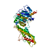
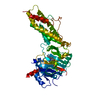
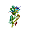
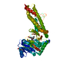
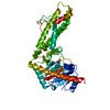
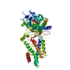
| Title | Crystal structure of the human fat mass and obesity associated protein (FTO) in complex with 5-carboxy-8-hydroxyquinoline (IOX1) | ||||||
 Components Components | Alpha-ketoglutarate-dependent dioxygenase FTO | ||||||
 Keywords Keywords | OXIDOREDUCTASE/OXIDOREDUCTASE INHIBITOR / double-stranded beta helix / jelly-roll motif / oxidoreductase / dioxygenase / nucleic acid demethylase / OXIDOREDUCTASE-OXIDOREDUCTASE INHIBITOR complex | ||||||
| Function / homology |  Function and homology information Function and homology informationregulation of white fat cell proliferation / tRNA demethylase activity / Reversal of alkylation damage by DNA dioxygenases / mRNA N6-methyladenine demethylase / mRNA N6-methyladenosine dioxygenase activity / regulation of respiratory system process / regulation of brown fat cell differentiation / regulation of lipid storage / broad specificity oxidative DNA demethylase activity / oxidative RNA demethylase activity ...regulation of white fat cell proliferation / tRNA demethylase activity / Reversal of alkylation damage by DNA dioxygenases / mRNA N6-methyladenine demethylase / mRNA N6-methyladenosine dioxygenase activity / regulation of respiratory system process / regulation of brown fat cell differentiation / regulation of lipid storage / broad specificity oxidative DNA demethylase activity / oxidative RNA demethylase activity / snRNA processing / RNA repair / DNA alkylation repair / Oxidoreductases; Acting on paired donors, with incorporation or reduction of molecular oxygen; With 2-oxoglutarate as one donor, and incorporation of one atom of oxygen into each donor / adipose tissue development / mRNA destabilization / regulation of multicellular organism growth / temperature homeostasis / ferrous iron binding / transferase activity / nuclear speck / nucleoplasm / nucleus / cytosol / cytoplasm Similarity search - Function | ||||||
| Biological species |  Homo sapiens (human) Homo sapiens (human) | ||||||
| Method |  X-RAY DIFFRACTION / MOLECULAR REPLACEMENT / molecular replacement / Resolution: 2.5048 Å X-RAY DIFFRACTION / MOLECULAR REPLACEMENT / molecular replacement / Resolution: 2.5048 Å | ||||||
 Authors Authors | Aik, W.S. / McDonough, M.A. / Schofield, C.J. | ||||||
 Citation Citation | Journal: J.Med.Chem. / Year: 2013 Title: Structural basis for inhibition of the fat mass and obesity associated protein (FTO) Authors: Aik, W.S. / Demetriades, M. / Hamdan, M.K.K. / Bagg, E.A.L. / Yeoh, K.K. / Lejeune, C. / Zhang, Z. / McDonough, M.A. / Schofield, C.J. | ||||||
| History |
|
- Structure visualization
| Structure viewer | Molecule: MolmilJmol/JSmol |
|---|
- Downloads & links
-Download
| PDBx/mmCIF format | 4ie4.cif.gz | 192.3 KB | Display | PDBx/mmCIF format |
|---|---|---|---|---|
| PDB format | pdb4ie4.ent.gz | 151 KB | Display | PDB format |
| PDBx/mmJSON format | 4ie4.json.gz | Tree view | PDBx/mmJSON format | |
| Others |  Other downloads Other downloads |
-Validation report
| Arichive directory | https://data.pdbj.org/pub/pdb/validation_reports/ie/4ie4ftp://data.pdbj.org/pub/pdb/validation_reports/ie/4ie4 | HTTPS FTP |
|---|
-Related structure data
| Related structure data |  4idzC  4ie0C 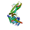 4ie5C 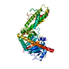 4ie6C 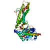 4ie7C 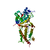 3lfmS S: Starting model for refinement C: citing same article ( |
|---|---|
| Similar structure data |


-Links
 PDBj
PDBj- Assembly
| Deposited unit | 
| ||||||||
|---|---|---|---|---|---|---|---|---|---|
| 1 |
| ||||||||
| Unit cell |
|
-Components
| #1: Protein | Mass: 56823.047 Da / Num. of mol.: 1 / Fragment: UNP residues 32-505 Source method: isolated from a genetically manipulated source Source: (gene. exp.) Homo sapiens (human) / Gene: FTO, KIAA1752 / Plasmid: pET28a / Production host:  References: UniProt: Q9C0B1, Oxidoreductases; Acting on paired donors, with incorporation or reduction of molecular oxygen; With 2-oxoglutarate as one donor, and incorporation of one atom of oxygen into each donor |
|---|---|
| #2: Chemical | ChemComp-ZN /   Mass: 65.409 Da / Num. of mol.: 1 / Source method: obtained synthetically / Formula: Zn Mass: 65.409 Da / Num. of mol.: 1 / Source method: obtained synthetically / Formula: Zn |
| #3: Chemical | ChemComp-GOL /   Mass: 92.094 Da / Num. of mol.: 1 / Source method: obtained synthetically / Formula: C3H8O3 Mass: 92.094 Da / Num. of mol.: 1 / Source method: obtained synthetically / Formula: C3H8O3 |
| #4: Chemical | ChemComp-8XQ /   Mass: 189.167 Da / Num. of mol.: 1 / Source method: obtained synthetically / Formula: C10H7NO3 Mass: 189.167 Da / Num. of mol.: 1 / Source method: obtained synthetically / Formula: C10H7NO3 |
| #5: Water | ChemComp-HOH /  Mass: 18.015 Da / Num. of mol.: 71 / Source method: isolated from a natural source / Formula: H2O Mass: 18.015 Da / Num. of mol.: 71 / Source method: isolated from a natural source / Formula: H2O |
-Experimental details
-Experiment
| Experiment | Method: X-RAY DIFFRACTION / Number of used crystals: 1 |
|---|
- Sample preparation
| Crystal | Density Matthews: 2.87 Å3/Da / Density % sol: 57.2 % / Mosaicity: 0.946 ° |
|---|---|
| Crystal grow | Temperature: 293 K / Method: vapor diffusion, hanging drop / pH: 5.6 Details: 13% PEG 3350, 100mM trisodium citrate pH 5.6, 4% tert-butanol, 1mM ZnSO4, 2mM 8XQ , VAPOR DIFFUSION, HANGING DROP, temperature 293K |
-Data collection
| Diffraction | Mean temperature: 100 K | |||||||||||||||||||||||||||||||||||||||||||||||||||||||||||||||||||||||||||||
|---|---|---|---|---|---|---|---|---|---|---|---|---|---|---|---|---|---|---|---|---|---|---|---|---|---|---|---|---|---|---|---|---|---|---|---|---|---|---|---|---|---|---|---|---|---|---|---|---|---|---|---|---|---|---|---|---|---|---|---|---|---|---|---|---|---|---|---|---|---|---|---|---|---|---|---|---|---|---|
| Diffraction source | Source: ROTATING ANODE / Type: RIGAKU FR-E+ SUPERBRIGHT / Wavelength: 1.5418 Å | |||||||||||||||||||||||||||||||||||||||||||||||||||||||||||||||||||||||||||||
| Detector | Type: RIGAKU SATURN 944+ / Detector: CCD / Date: Nov 15, 2012 / Details: osmic HF | |||||||||||||||||||||||||||||||||||||||||||||||||||||||||||||||||||||||||||||
| Radiation | Protocol: SINGLE WAVELENGTH / Monochromatic (M) / Laue (L): M / Scattering type: x-ray | |||||||||||||||||||||||||||||||||||||||||||||||||||||||||||||||||||||||||||||
| Radiation wavelength | Wavelength: 1.5418 Å / Relative weight: 1 | |||||||||||||||||||||||||||||||||||||||||||||||||||||||||||||||||||||||||||||
| Reflection | Resolution: 2.5→50 Å / Num. all: 21755 / Num. obs: 21717 / % possible obs: 99.8 % / Observed criterion σ(I): -3 / Redundancy: 4.9 % / Biso Wilson estimate: 52.28 Å2 / Rmerge(I) obs: 0.177 / Χ2: 1.071 / Net I/σ(I): 12.9 | |||||||||||||||||||||||||||||||||||||||||||||||||||||||||||||||||||||||||||||
| Reflection shell | Diffraction-ID: 1
|
-Phasing
| Phasing | Method: molecular replacement | |||||||||
|---|---|---|---|---|---|---|---|---|---|---|
| Phasing MR |
|
- Processing
| Software |
| ||||||||||||||||||||||||||||||||||||
|---|---|---|---|---|---|---|---|---|---|---|---|---|---|---|---|---|---|---|---|---|---|---|---|---|---|---|---|---|---|---|---|---|---|---|---|---|---|
| Refinement | Method to determine structure: MOLECULAR REPLACEMENT Starting model: PDB entry 3LFM Resolution: 2.5048→28.8308 Å / Occupancy max: 1 / Occupancy min: 0.5 / Isotropic thermal model: Isotropic
| ||||||||||||||||||||||||||||||||||||
| Displacement parameters | Biso max: 125.5 Å2 / Biso mean: 47.8656 Å2 / Biso min: 3.07 Å2 | ||||||||||||||||||||||||||||||||||||
| Refinement step | Cycle: LAST / Resolution: 2.5048→28.8308 Å
| ||||||||||||||||||||||||||||||||||||
| Refine LS restraints |
| ||||||||||||||||||||||||||||||||||||
| LS refinement shell |
|
