 Movie
Movie Controller

[English] 日本語
 Yorodumi
- PDB-4i7a: GrpN pentameric microcompartment shell protein from Rhodospirillu... -
+ Open data

- Basic information
| Entry | Database: PDB / ID: 4i7a | ||||||
|---|---|---|---|---|---|---|---|
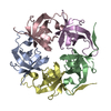
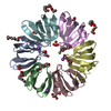
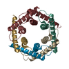
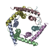
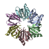
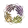
| Title | GrpN pentameric microcompartment shell protein from Rhodospirillum rubrum | ||||||
 Components Components | Ethanolamine utilization protein EutN/carboxysome structural protein Ccml | ||||||
 Keywords Keywords | STRUCTURAL PROTEIN / microcompartments / ccmL / eutN / glycyl radical propanediol microcompartment | ||||||
| Function / homology | EutN/Ccml / OB fold (Dihydrolipoamide Acetyltransferase, E2P) / Beta Barrel / Mainly Beta / :  Function and homology information Function and homology information | ||||||
| Biological species |  Rhodospirillum rubrum (bacteria) Rhodospirillum rubrum (bacteria) | ||||||
| Method |  X-RAY DIFFRACTION / SYNCHROTRON / MOLECULAR REPLACEMENT / molecular replacement / Resolution: 3.2 Å X-RAY DIFFRACTION / SYNCHROTRON / MOLECULAR REPLACEMENT / molecular replacement / Resolution: 3.2 Å | ||||||
 Authors Authors | Wheatley, N.M. / Gidaniyan, S.D. / Cascio, D. / Yeates, T.O. | ||||||
 Citation Citation | Journal: Protein Sci. / Year: 2013 Title: Bacterial microcompartment shells of diverse functional types possess pentameric vertex proteins. Authors: Wheatley, N.M. / Gidaniyan, S.D. / Liu, Y. / Cascio, D. / Yeates, T.O. | ||||||
| History |
|
- Structure visualization
| Structure viewer | Molecule: MolmilJmol/JSmol |
|---|
- Downloads & links
-Download
| PDBx/mmCIF format | 4i7a.cif.gz | 158.2 KB | Display | PDBx/mmCIF format |
|---|---|---|---|---|
| PDB format | pdb4i7a.ent.gz | 127.5 KB | Display | PDB format |
| PDBx/mmJSON format | 4i7a.json.gz | Tree view | PDBx/mmJSON format | |
| Others |  Other downloads Other downloads |
-Validation report
| Arichive directory | https://data.pdbj.org/pub/pdb/validation_reports/i7/4i7aftp://data.pdbj.org/pub/pdb/validation_reports/i7/4i7a | HTTPS FTP |
|---|
-Related structure data
| Related structure data |  2qw7S S: Starting model for refinement |
|---|---|
| Similar structure data |


-Links
 PDBj
PDBj- Assembly
| Deposited unit | 
| ||||||||
|---|---|---|---|---|---|---|---|---|---|
| 1 |
| ||||||||
| Unit cell |
|
-Components
| #1: Protein | Mass: 9880.153 Da / Num. of mol.: 5 Source method: isolated from a genetically manipulated source Source: (gene. exp.) Rhodospirillum rubrum (bacteria) / Strain: F11 / Gene: F11_04705, grpN / Plasmid: pET22b(+) / Production host: #2: Chemical |   Mass: 35.453 Da / Num. of mol.: 3 / Source method: obtained synthetically / Formula: Cl Mass: 35.453 Da / Num. of mol.: 3 / Source method: obtained synthetically / Formula: Cl#3: Water | ChemComp-HOH / |  Mass: 18.015 Da / Num. of mol.: 1 / Source method: isolated from a natural source / Formula: H2O Mass: 18.015 Da / Num. of mol.: 1 / Source method: isolated from a natural source / Formula: H2O |
|---|
-Experimental details
-Experiment
| Experiment | Method: X-RAY DIFFRACTION / Number of used crystals: 1 |
|---|
- Sample preparation
| Crystal | Density Matthews: 2.89 Å3/Da / Density % sol: 57.4 % |
|---|---|
| Crystal grow | Temperature: 298 K / Method: vapor diffusion, hanging drop / pH: 7.2 Details: 0.1 M sodium chloride, 0.1 M HEPES, pH 7.2, 1.5 M ammonium sulfate, VAPOR DIFFUSION, HANGING DROP, temperature 298K |
-Data collection
| Diffraction | Mean temperature: 100 K | |||||||||||||||||||||||||||||||||||||||||||||||||||||||||||||||||||||||||||||||||||||||||||||||||||||||||||||||||||||||||||||||||||||||||||||||||||
|---|---|---|---|---|---|---|---|---|---|---|---|---|---|---|---|---|---|---|---|---|---|---|---|---|---|---|---|---|---|---|---|---|---|---|---|---|---|---|---|---|---|---|---|---|---|---|---|---|---|---|---|---|---|---|---|---|---|---|---|---|---|---|---|---|---|---|---|---|---|---|---|---|---|---|---|---|---|---|---|---|---|---|---|---|---|---|---|---|---|---|---|---|---|---|---|---|---|---|---|---|---|---|---|---|---|---|---|---|---|---|---|---|---|---|---|---|---|---|---|---|---|---|---|---|---|---|---|---|---|---|---|---|---|---|---|---|---|---|---|---|---|---|---|---|---|---|---|---|
| Diffraction source | Source: SYNCHROTRON / Site: APS  / Beamline: 24-ID-C / Wavelength: 0.9791 Å / Beamline: 24-ID-C / Wavelength: 0.9791 Å | |||||||||||||||||||||||||||||||||||||||||||||||||||||||||||||||||||||||||||||||||||||||||||||||||||||||||||||||||||||||||||||||||||||||||||||||||||
| Detector | Type: DECTRIS PILATUS 6M / Detector: PIXEL / Date: Oct 27, 2012 | |||||||||||||||||||||||||||||||||||||||||||||||||||||||||||||||||||||||||||||||||||||||||||||||||||||||||||||||||||||||||||||||||||||||||||||||||||
| Radiation | Monochromator: double crystal Si(111) / Protocol: SINGLE WAVELENGTH / Monochromatic (M) / Laue (L): M / Scattering type: x-ray | |||||||||||||||||||||||||||||||||||||||||||||||||||||||||||||||||||||||||||||||||||||||||||||||||||||||||||||||||||||||||||||||||||||||||||||||||||
| Radiation wavelength | Wavelength: 0.9791 Å / Relative weight: 1 | |||||||||||||||||||||||||||||||||||||||||||||||||||||||||||||||||||||||||||||||||||||||||||||||||||||||||||||||||||||||||||||||||||||||||||||||||||
| Reflection | Resolution: 3.2→106.57 Å / Num. obs: 9248 / % possible obs: 96.6 % / Observed criterion σ(I): -3 / Redundancy: 5.4 % / Biso Wilson estimate: 73.556 Å2 / Rmerge(I) obs: 0.066 / Net I/σ(I): 21.02 | |||||||||||||||||||||||||||||||||||||||||||||||||||||||||||||||||||||||||||||||||||||||||||||||||||||||||||||||||||||||||||||||||||||||||||||||||||
| Reflection shell |
|
-Phasing
| Phasing | Method: molecular replacement | |||||||||
|---|---|---|---|---|---|---|---|---|---|---|
| Phasing MR | Model details: Phaser MODE: MR_AUTO
|
- Processing
| Software |
| ||||||||||||||||||||||||||||||||||||||||
|---|---|---|---|---|---|---|---|---|---|---|---|---|---|---|---|---|---|---|---|---|---|---|---|---|---|---|---|---|---|---|---|---|---|---|---|---|---|---|---|---|---|
| Refinement | Method to determine structure: MOLECULAR REPLACEMENT Starting model: PDB ENTRY 2QW7 Resolution: 3.2→19.789 Å / Occupancy max: 1 / Occupancy min: 1 / FOM work R set: 0.7272 / SU ML: 0.39 / Cross valid method: THROUGHOUT / σ(F): 1.99 / Phase error: 33 / Stereochemistry target values: ML
| ||||||||||||||||||||||||||||||||||||||||
| Solvent computation | Shrinkage radii: 0.9 Å / VDW probe radii: 1.11 Å / Solvent model: FLAT BULK SOLVENT MODEL | ||||||||||||||||||||||||||||||||||||||||
| Displacement parameters | Biso max: 160.25 Å2 / Biso mean: 87.6177 Å2 / Biso min: 51.97 Å2 | ||||||||||||||||||||||||||||||||||||||||
| Refinement step | Cycle: LAST / Resolution: 3.2→19.789 Å
| ||||||||||||||||||||||||||||||||||||||||
| Refine LS restraints |
| ||||||||||||||||||||||||||||||||||||||||
| LS refinement shell | Refine-ID: X-RAY DIFFRACTION / Total num. of bins used: 3
| ||||||||||||||||||||||||||||||||||||||||
| Refinement TLS params. | Method: refined / Origin x: 47.2819 Å / Origin y: 29.815 Å / Origin z: 0.0653 Å
| ||||||||||||||||||||||||||||||||||||||||
| Refinement TLS group |
|
