 Movie
Movie Controller


+ Open data

- Basic information
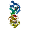
| Entry | Database: PDB / ID: 4i66 | ||||||
|---|---|---|---|---|---|---|---|
| Title | Crystal structure of Hoch_4089 protein from Haliangium ochraceum | ||||||
 Components Components | uncharacterized protein Hoch_4089 | ||||||
 Keywords Keywords | Structural Genomics / Unknown Function / PSI-Biology / Midwest Center for Structural Genomics / MCSG | ||||||
| Function / homology | Domain of unknown function DUF4159 / Domain of unknown function DUF4159 / Domain of unknown function (DUF4159) / Twin arginine translocation (Tat) signal profile. / Twin-arginine translocation pathway, signal sequence / Rossmann fold / 3-Layer(aba) Sandwich / Alpha Beta / DUF4159 domain-containing protein Function and homology information Function and homology information | ||||||
| Biological species |  Haliangium ochraceum (bacteria) Haliangium ochraceum (bacteria) | ||||||
| Method |  X-RAY DIFFRACTION / SYNCHROTRON / SAD / Resolution: 1.3 Å X-RAY DIFFRACTION / SYNCHROTRON / SAD / Resolution: 1.3 Å | ||||||
 Authors Authors | Michalska, K. / Mack, J.C. / Endres, M. / Joachimiak, A. / Midwest Center for Structural Genomics (MCSG) | ||||||
 Citation Citation | Journal: To be Published Title: Crystal structure of Hoch_4089 protein from Haliangium ochraceum Authors: Michalska, K. / Mack, J.C. / Endres, M. / Joachimiak, A. | ||||||
| History |
|
- Structure visualization
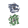
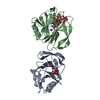
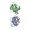
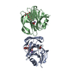
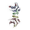
| Structure viewer | Molecule: MolmilJmol/JSmol |
|---|
- Downloads & links
-Download
| PDBx/mmCIF format | 4i66.cif.gz | 99.3 KB | Display | PDBx/mmCIF format |
|---|---|---|---|---|
| PDB format | pdb4i66.ent.gz | 76 KB | Display | PDB format |
| PDBx/mmJSON format | 4i66.json.gz | Tree view | PDBx/mmJSON format | |
| Others |  Other downloads Other downloads |
-Validation report
| Arichive directory | https://data.pdbj.org/pub/pdb/validation_reports/i6/4i66ftp://data.pdbj.org/pub/pdb/validation_reports/i6/4i66 | HTTPS FTP |
|---|
-Related structure data
| Similar structure data | |
|---|---|
| Other databases |


-Links
 PDBj
PDBj

- Assembly
| Deposited unit | 
| |||||||||
|---|---|---|---|---|---|---|---|---|---|---|
| 1 | 
| |||||||||
| Unit cell |
| |||||||||
| Components on special symmetry positions |
|
-Components
| #1: Protein | Mass: 23387.984 Da / Num. of mol.: 1 Source method: isolated from a genetically manipulated source Source: (gene. exp.) Haliangium ochraceum (bacteria) / Strain: DSM 14365 / Gene: Hoch_4089 / Plasmid: pMCSG68 / Production host: | ||||
|---|---|---|---|---|---|
| #2: Chemical |   Mass: 96.063 Da / Num. of mol.: 2 / Source method: obtained synthetically / Formula: SO4 Mass: 96.063 Da / Num. of mol.: 2 / Source method: obtained synthetically / Formula: SO4#3: Water | ChemComp-HOH / |  Mass: 18.015 Da / Num. of mol.: 174 / Source method: isolated from a natural source / Formula: H2O Mass: 18.015 Da / Num. of mol.: 174 / Source method: isolated from a natural source / Formula: H2OHas protein modification | Y | |
-Experimental details
-Experiment
| Experiment | Method: X-RAY DIFFRACTION / Number of used crystals: 1 |
|---|
- Sample preparation
| Crystal | Density Matthews: 1.98 Å3/Da / Density % sol: 37.89 % |
|---|---|
| Crystal grow | Temperature: 297 K / Method: vapor diffusion, sitting drop / pH: 7.5 Details: 0.2 M Li2SO4, 0.1 M HEPES/NaOH pH 7.5, 25% PEG3350, VAPOR DIFFUSION, SITTING DROP, temperature 297K |
-Data collection
| Diffraction | Mean temperature: 100 K |
|---|---|
| Diffraction source | Source: SYNCHROTRON / Site: APS  / Beamline: 19-ID / Wavelength: 0.97915 Å / Beamline: 19-ID / Wavelength: 0.97915 Å |
| Detector | Type: ADSC QUANTUM 315r / Detector: CCD / Date: Oct 22, 2012 / Details: mirrors |
| Radiation | Monochromator: double crystal / Protocol: SINGLE WAVELENGTH / Monochromatic (M) / Laue (L): M / Scattering type: x-ray |
| Radiation wavelength | Wavelength: 0.97915 Å / Relative weight: 1 |
| Reflection | Resolution: 1.3→30 Å / Num. all: 46523 / Num. obs: 46401 / % possible obs: 99.7 % / Observed criterion σ(I): -3 / Redundancy: 9 % / Biso Wilson estimate: 14 Å2 / Rmerge(I) obs: 0.086 / Net I/σ(I): 30.5 |
| Reflection shell | Resolution: 1.3→1.32 Å / Redundancy: 8.9 % / Rmerge(I) obs: 0.848 / Mean I/σ(I) obs: 2.7 / Num. unique all: 2297 / % possible all: 100 |
- Processing
| Software |
| ||||||||||||||||||||||||||||||||||||||||||||||||||||||||||||||||||||||||||||||||||||||||||
|---|---|---|---|---|---|---|---|---|---|---|---|---|---|---|---|---|---|---|---|---|---|---|---|---|---|---|---|---|---|---|---|---|---|---|---|---|---|---|---|---|---|---|---|---|---|---|---|---|---|---|---|---|---|---|---|---|---|---|---|---|---|---|---|---|---|---|---|---|---|---|---|---|---|---|---|---|---|---|---|---|---|---|---|---|---|---|---|---|---|---|---|
| Refinement | Method to determine structure: SAD / Resolution: 1.3→22.25 Å / Cor.coef. Fo:Fc: 0.97 / Cor.coef. Fo:Fc free: 0.953 / SU B: 1.584 / SU ML: 0.031 / Isotropic thermal model: anisotropic / Cross valid method: THROUGHOUT / ESU R: 0.052 / ESU R Free: 0.052 / Stereochemistry target values: MAXIMUM LIKELIHOOD / Details: HYDROGENS HAVE BEEN ADDED IN THE RIDING POSITIONS
| ||||||||||||||||||||||||||||||||||||||||||||||||||||||||||||||||||||||||||||||||||||||||||
| Solvent computation | Ion probe radii: 0.8 Å / Shrinkage radii: 0.8 Å / VDW probe radii: 1.4 Å / Solvent model: MASK | ||||||||||||||||||||||||||||||||||||||||||||||||||||||||||||||||||||||||||||||||||||||||||
| Displacement parameters | Biso mean: 14.766 Å2
| ||||||||||||||||||||||||||||||||||||||||||||||||||||||||||||||||||||||||||||||||||||||||||
| Refinement step | Cycle: LAST / Resolution: 1.3→22.25 Å
| ||||||||||||||||||||||||||||||||||||||||||||||||||||||||||||||||||||||||||||||||||||||||||
| Refine LS restraints |
| ||||||||||||||||||||||||||||||||||||||||||||||||||||||||||||||||||||||||||||||||||||||||||
| LS refinement shell | Resolution: 1.299→1.333 Å / Total num. of bins used: 20
|
