 Movie
Movie Controller

[English] 日本語
 Yorodumi
- PDB-4hwm: Crystal structure of a lipoprotein YedD (KPN_02420) from Klebsiel... -
+ Open data

- Basic information
| Entry | Database: PDB / ID: 4hwm | ||||||
|---|---|---|---|---|---|---|---|
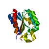
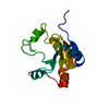
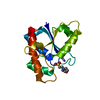
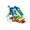
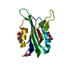
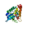
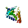
| Title | Crystal structure of a lipoprotein YedD (KPN_02420) from Klebsiella pneumoniae subsp. pneumoniae MGH 78578 at 1.38 A resolution | ||||||
 Components Components | Uncharacterized protein yedD | ||||||
 Keywords Keywords | STRUCTURAL GENOMICS / UNKNOWN FUNCTION / YedD-like protein / PF13987 family / Joint Center for Structural Genomics / JCSG / Protein Structure Initiative / PSI-BIOLOGY | ||||||
| Function / homology | YedD-like protein / YedD-like protein / YedD-like superfamily / Bacterial lipocalin LPS transport optimiser YedD / Lipocalin / Prokaryotic membrane lipoprotein lipid attachment site profile. / Beta Barrel / Mainly Beta / Lipoprotein Function and homology information Function and homology information | ||||||
| Biological species |  Klebsiella pneumoniae subsp. pneumoniae (bacteria) Klebsiella pneumoniae subsp. pneumoniae (bacteria) | ||||||
| Method |  X-RAY DIFFRACTION / SYNCHROTRON / SAD / Resolution: 1.38 Å X-RAY DIFFRACTION / SYNCHROTRON / SAD / Resolution: 1.38 Å | ||||||
 Authors Authors | Joint Center for Structural Genomics (JCSG) | ||||||
 Citation Citation | Journal: To be published Title: Crystal structure of a lipoprotein YedD (KPN_02420) from Klebsiella pneumoniae subsp. pneumoniae MGH 78578 at 1.38 A resolution Authors: Joint Center for Structural Genomics (JCSG) | ||||||
| History |
|
- Structure visualization
| Structure viewer | Molecule: MolmilJmol/JSmol |
|---|
- Downloads & links
-Download
| PDBx/mmCIF format | 4hwm.cif.gz | 70.2 KB | Display | PDBx/mmCIF format |
|---|---|---|---|---|
| PDB format | pdb4hwm.ent.gz | 51.9 KB | Display | PDB format |
| PDBx/mmJSON format | 4hwm.json.gz | Tree view | PDBx/mmJSON format | |
| Others |  Other downloads Other downloads |
-Validation report
| Arichive directory | https://data.pdbj.org/pub/pdb/validation_reports/hw/4hwmftp://data.pdbj.org/pub/pdb/validation_reports/hw/4hwm | HTTPS FTP |
|---|
-Related structure data
| Similar structure data | |
|---|---|
| Other databases |


-Links
 PDBj
PDBj


- Assembly
| Deposited unit | 
| ||||||||
|---|---|---|---|---|---|---|---|---|---|
| 1 |
| ||||||||
| Unit cell |
|
-Components
| #1: Protein | Mass: 14453.691 Da / Num. of mol.: 1 / Fragment: UNP residues 16-144 Source method: isolated from a genetically manipulated source Source: (gene. exp.) Klebsiella pneumoniae subsp. pneumoniae (bacteria)Strain: ATCC 700721 / MGH 78578 / Gene: yedD, KPN78578_23820, KPN_02420 / Plasmid: SpeedET / Production host: |
|---|---|
| #2: Water | ChemComp-HOH /  Mass: 18.015 Da / Num. of mol.: 154 / Source method: isolated from a natural source / Formula: H2O Mass: 18.015 Da / Num. of mol.: 154 / Source method: isolated from a natural source / Formula: H2O |
| Has protein modification | Y |
| Sequence details | THE CONSTRUCT (16-144) WAS EXPRESSED WITH A PURIFICATION TAG MGSDKIHHHHHHENLYFQG. THE TAG WAS ...THE CONSTRUCT (16-144) WAS EXPRESSED WITH A PURIFICATI |
-Experimental details
-Experiment
| Experiment | Method: X-RAY DIFFRACTION / Number of used crystals: 1 |
|---|
- Sample preparation
| Crystal | Density Matthews: 1.84 Å3/Da / Density % sol: 33.29 % |
|---|---|
| Crystal grow | Temperature: 293 K / Method: vapor diffusion, sitting drop / pH: 9.5 Details: 30.00% polyethylene glycol 3000, 0.1M CHES pH 9.5, NANODROP, VAPOR DIFFUSION, SITTING DROP, temperature 293K |
-Data collection
| Diffraction | Mean temperature: 100 K | |||||||||||||||||||||||||||||||||||||||||||||||||||||||||||||||||||||||||||||
|---|---|---|---|---|---|---|---|---|---|---|---|---|---|---|---|---|---|---|---|---|---|---|---|---|---|---|---|---|---|---|---|---|---|---|---|---|---|---|---|---|---|---|---|---|---|---|---|---|---|---|---|---|---|---|---|---|---|---|---|---|---|---|---|---|---|---|---|---|---|---|---|---|---|---|---|---|---|---|
| Diffraction source | Source: SYNCHROTRON / Site: ALS  / Beamline: 8.2.2 / Wavelength: 0.979133 / Beamline: 8.2.2 / Wavelength: 0.979133 | |||||||||||||||||||||||||||||||||||||||||||||||||||||||||||||||||||||||||||||
| Detector | Type: ADSC QUANTUM 315 / Detector: CCD / Date: Sep 19, 2012 / Details: KOHZU: Double Crystal Si(111) | |||||||||||||||||||||||||||||||||||||||||||||||||||||||||||||||||||||||||||||
| Radiation | Monochromator: Double Crystal Si(111) / Protocol: SINGLE WAVELENGTH / Monochromatic (M) / Laue (L): M / Scattering type: x-ray | |||||||||||||||||||||||||||||||||||||||||||||||||||||||||||||||||||||||||||||
| Radiation wavelength | Wavelength: 0.979133 Å / Relative weight: 1 | |||||||||||||||||||||||||||||||||||||||||||||||||||||||||||||||||||||||||||||
| Reflection | Resolution: 1.38→39.81 Å / Num. obs: 22613 / % possible obs: 99.8 % / Observed criterion σ(I): -3 / Redundancy: 7 % / Biso Wilson estimate: 16.745 Å2 / Rmerge(I) obs: 0.045 / Net I/σ(I): 15.44 | |||||||||||||||||||||||||||||||||||||||||||||||||||||||||||||||||||||||||||||
| Reflection shell |
|
-Phasing
| Phasing | Method: SAD |
|---|
- Processing
| Software |
| ||||||||||||||||||||||||||||||||||||||||||||||||||||||||||||||||||||||||||||||||||||||||||||||||||||
|---|---|---|---|---|---|---|---|---|---|---|---|---|---|---|---|---|---|---|---|---|---|---|---|---|---|---|---|---|---|---|---|---|---|---|---|---|---|---|---|---|---|---|---|---|---|---|---|---|---|---|---|---|---|---|---|---|---|---|---|---|---|---|---|---|---|---|---|---|---|---|---|---|---|---|---|---|---|---|---|---|---|---|---|---|---|---|---|---|---|---|---|---|---|---|---|---|---|---|---|---|---|
| Refinement | Method to determine structure: SAD / Resolution: 1.38→39.81 Å / Cor.coef. Fo:Fc: 0.974 / Cor.coef. Fo:Fc free: 0.969 / Occupancy max: 1 / Occupancy min: 0.15 / SU B: 2.145 / SU ML: 0.038 / Cross valid method: THROUGHOUT / σ(F): 0 / ESU R: 0.068 / ESU R Free: 0.059 Stereochemistry target values: MAXIMUM LIKELIHOOD WITH PHASES Details: 1. A MET-INHIBITION PROTOCOL WAS USED FOR SELENOMETHIONINE INCORPORATION DURING PROTEIN EXPRESSION. THE OCCUPANCY OF THE SE ATOMS IN THE MSE RESIDUES WAS REDUCED TO 0.75 FOR THE REDUCED ...Details: 1. A MET-INHIBITION PROTOCOL WAS USED FOR SELENOMETHIONINE INCORPORATION DURING PROTEIN EXPRESSION. THE OCCUPANCY OF THE SE ATOMS IN THE MSE RESIDUES WAS REDUCED TO 0.75 FOR THE REDUCED SCATTERING POWER DUE TO PARTIAL S-MET INCORPORATION. 2. HYDROGENS HAVE BEEN ADDED IN THE RIDING POSITIONS.
| ||||||||||||||||||||||||||||||||||||||||||||||||||||||||||||||||||||||||||||||||||||||||||||||||||||
| Solvent computation | Ion probe radii: 0.8 Å / Shrinkage radii: 0.8 Å / VDW probe radii: 1.2 Å / Solvent model: MASK | ||||||||||||||||||||||||||||||||||||||||||||||||||||||||||||||||||||||||||||||||||||||||||||||||||||
| Displacement parameters | Biso max: 64.57 Å2 / Biso mean: 21.1093 Å2 / Biso min: 9.32 Å2
| ||||||||||||||||||||||||||||||||||||||||||||||||||||||||||||||||||||||||||||||||||||||||||||||||||||
| Refinement step | Cycle: LAST / Resolution: 1.38→39.81 Å
| ||||||||||||||||||||||||||||||||||||||||||||||||||||||||||||||||||||||||||||||||||||||||||||||||||||
| Refine LS restraints |
| ||||||||||||||||||||||||||||||||||||||||||||||||||||||||||||||||||||||||||||||||||||||||||||||||||||
| LS refinement shell | Resolution: 1.38→1.416 Å / Total num. of bins used: 20
|
