 Movie
Movie Controller

[English] 日本語
 Yorodumi
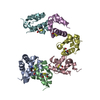
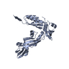
- PDB-4hpz: Crystal structure of a TALE protein reveals an extended N-termina... -
+ Open data

- Basic information
| Entry | Database: PDB / ID: 4hpz | ||||||
|---|---|---|---|---|---|---|---|
| Title | Crystal structure of a TALE protein reveals an extended N-terminal DNA binding region | ||||||
 Components Components | dTale2 | ||||||
 Keywords Keywords | DNA BINDING PROTEIN / N-TERMINAL DOMAIN / TAL EFFECTOR | ||||||
| Function / homology | TAL effector repeat / TAL effector repeat / host cell nucleus / extracellular region / dTale2 Function and homology information Function and homology information | ||||||
| Biological species |  Xanthomonas (bacteria) Xanthomonas (bacteria) | ||||||
| Method |  X-RAY DIFFRACTION / SYNCHROTRON / MOLECULAR REPLACEMENT / Resolution: 2.202 Å X-RAY DIFFRACTION / SYNCHROTRON / MOLECULAR REPLACEMENT / Resolution: 2.202 Å | ||||||
 Authors Authors | Chai, J. / Han, Z. / Gao, H. | ||||||
 Citation Citation | Journal: Cell Res. / Year: 2012 Title: Crystal structure of a TALE protein reveals an extended N-terminal DNA binding region Authors: Gao, H. / Wu, X. / Chai, J. / Han, Z. | ||||||
| History |
|
- Structure visualization
| Structure viewer | Molecule: MolmilJmol/JSmol |
|---|
- Downloads & links
-Download
| PDBx/mmCIF format | 4hpz.cif.gz | 172.3 KB | Display | PDBx/mmCIF format |
|---|---|---|---|---|
| PDB format | pdb4hpz.ent.gz | 136.8 KB | Display | PDB format |
| PDBx/mmJSON format | 4hpz.json.gz | Tree view | PDBx/mmJSON format | |
| Others |  Other downloads Other downloads |
-Validation report
| Summary document | 4hpz_validation.pdf.gz | 439 KB | Display | wwPDB validaton report |
|---|---|---|---|---|
| Full document | 4hpz_full_validation.pdf.gz | 450.4 KB | Display | |
| Data in XML | 4hpz_validation.xml.gz | 19.4 KB | Display | |
| Data in CIF | 4hpz_validation.cif.gz | 27.7 KB | Display | |
| Arichive directory | https://data.pdbj.org/pub/pdb/validation_reports/hp/4hpzftp://data.pdbj.org/pub/pdb/validation_reports/hp/4hpz | HTTPS FTP |
-Related structure data
| Related structure data | 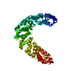 3v6pS S: Starting model for refinement |
|---|---|
| Similar structure data |


-Links
 PDBj
PDBj
- Assembly
| Deposited unit | 
| ||||||||
|---|---|---|---|---|---|---|---|---|---|
| 1 |
| ||||||||
| Unit cell |
| ||||||||
| Components on special symmetry positions |
|
-Components
| #1: Protein | Mass: 48280.113 Da / Num. of mol.: 1 / Fragment: dTale2 Source method: isolated from a genetically manipulated source Source: (gene. exp.) Xanthomonas (bacteria) / Plasmid: pGEX-6P-1 / Production host: | ||||
|---|---|---|---|---|---|
| #2: Chemical |   Mass: 96.063 Da / Num. of mol.: 3 / Source method: obtained synthetically / Formula: SO4 Mass: 96.063 Da / Num. of mol.: 3 / Source method: obtained synthetically / Formula: SO4#3: Water | ChemComp-HOH / |  Mass: 18.015 Da / Num. of mol.: 160 / Source method: isolated from a natural source / Formula: H2O Mass: 18.015 Da / Num. of mol.: 160 / Source method: isolated from a natural source / Formula: H2OSequence details | A SEQUENCE DATABASE REFERENCE FOR THIS PROTEIN DOES NOT CURRENTLY EXIST. THIS HIGHLY-CONSERVED ...A SEQUENCE DATABASE REFERENCE FOR THIS PROTEIN DOES NOT CURRENTLY EXIST. THIS HIGHLY-CONSERVED PROTEIN WAS CONSTRUCTE | |
-Experimental details
-Experiment
| Experiment | Method: X-RAY DIFFRACTION / Number of used crystals: 1 |
|---|
- Sample preparation
| Crystal | Density Matthews: 2.93 Å3/Da / Density % sol: 58.04 % |
|---|---|
| Crystal grow | Temperature: 291 K / Method: vapor diffusion, hanging drop / pH: 7 Details: 0.05M MgSO4, 0.05M HEPES, 1.6M Li2SO4, pH 7.0, VAPOR DIFFUSION, HANGING DROP, temperature 291K |
-Data collection
| Diffraction | Mean temperature: 100 K |
|---|---|
| Diffraction source | Source: SYNCHROTRON / Site: SSRF  / Beamline: BL17U / Wavelength: 0.9791 Å / Beamline: BL17U / Wavelength: 0.9791 Å |
| Detector | Type: ADSC QUANTUM 315 / Detector: CCD / Date: Sep 27, 2011 |
| Radiation | Monochromator: SAGITALLY FOCUSED Si(111) / Protocol: SINGLE WAVELENGTH / Monochromatic (M) / Laue (L): M / Scattering type: x-ray |
| Radiation wavelength | Wavelength: 0.9791 Å / Relative weight: 1 |
| Reflection | Resolution: 2.2→15 Å / Num. all: 29024 / Num. obs: 29024 / % possible obs: 100 % / Observed criterion σ(F): 2 / Observed criterion σ(I): 2 / Redundancy: 10.9 % / Rmerge(I) obs: 0.069 |
| Reflection shell | Resolution: 2.2→2.24 Å / % possible all: 85.9 |
- Processing
| Software |
| |||||||||||||||||||||||||||||||||||||||||||||||||||||||
|---|---|---|---|---|---|---|---|---|---|---|---|---|---|---|---|---|---|---|---|---|---|---|---|---|---|---|---|---|---|---|---|---|---|---|---|---|---|---|---|---|---|---|---|---|---|---|---|---|---|---|---|---|---|---|---|---|
| Refinement | Method to determine structure: MOLECULAR REPLACEMENT Starting model: 3V6P Resolution: 2.202→14.975 Å / SU ML: 0.24 / σ(F): 0 / Phase error: 26.23 / Stereochemistry target values: ML
| |||||||||||||||||||||||||||||||||||||||||||||||||||||||
| Solvent computation | Shrinkage radii: 0.95 Å / VDW probe radii: 1.2 Å / Solvent model: FLAT BULK SOLVENT MODEL / Bsol: 40.732 Å2 / ksol: 0.392 e/Å3 | |||||||||||||||||||||||||||||||||||||||||||||||||||||||
| Displacement parameters |
| |||||||||||||||||||||||||||||||||||||||||||||||||||||||
| Refinement step | Cycle: LAST / Resolution: 2.202→14.975 Å
| |||||||||||||||||||||||||||||||||||||||||||||||||||||||
| Refine LS restraints |
| |||||||||||||||||||||||||||||||||||||||||||||||||||||||
| LS refinement shell | Refine-ID: X-RAY DIFFRACTION / Total num. of bins used: 10 / % reflection obs: 100 %
| |||||||||||||||||||||||||||||||||||||||||||||||||||||||
| Refinement TLS params. | Method: refined / Origin x: 17.7169 Å / Origin y: 55.0361 Å / Origin z: -25.2695 Å
| |||||||||||||||||||||||||||||||||||||||||||||||||||||||
| Refinement TLS group | Selection details: all |
