 Movie
Movie Controller

[English] 日本語
 Yorodumi
- PDB-4ho2: Crystal structure of glucose 1-phosphate thymidylyltransferase fr... -
+ Open data

- Basic information
| Entry | Database: PDB / ID: 4ho2 | ||||||
|---|---|---|---|---|---|---|---|
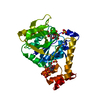
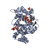
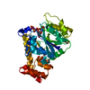
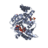
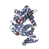
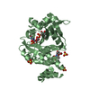
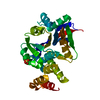
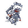
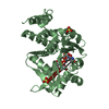
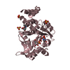
| Title | Crystal structure of glucose 1-phosphate thymidylyltransferase from Aneurinibacillus thermoaerophilus complexed with thymidine | ||||||
 Components Components | Glucose-1-phosphate thymidylyltransferase | ||||||
 Keywords Keywords | TRANSFERASE / thymidylyltransferase / nucleotide binding | ||||||
| Function / homology |  Function and homology information Function and homology informationglucose-1-phosphate thymidylyltransferase / glucose-1-phosphate thymidylyltransferase activity / biosynthetic process / nucleotide binding / metal ion binding Similarity search - Function | ||||||
| Biological species |  Aneurinibacillus thermoaerophilus (bacteria) Aneurinibacillus thermoaerophilus (bacteria) | ||||||
| Method |  X-RAY DIFFRACTION / SYNCHROTRON / MOLECULAR REPLACEMENT / Resolution: 1.84 Å X-RAY DIFFRACTION / SYNCHROTRON / MOLECULAR REPLACEMENT / Resolution: 1.84 Å | ||||||
 Authors Authors | Chen, T.J. / Chien, W.T. / Lin, C.C. / Wang, W.C. | ||||||
 Citation Citation | Journal: TO BE PUBLISHED Title: Crystal structure of glucose 1-phosphate thymidylyltransferase from Aneurinibacillus thermoaerophilus complexed with thymidine Authors: Chen, T.J. / Chien, W.T. / Lin, C.C. / Wang, W.C. | ||||||
| History |
|
- Structure visualization
| Structure viewer | Molecule: MolmilJmol/JSmol |
|---|
- Downloads & links
-Download
| PDBx/mmCIF format | 4ho2.cif.gz | 76.5 KB | Display | PDBx/mmCIF format |
|---|---|---|---|---|
| PDB format | pdb4ho2.ent.gz | 56 KB | Display | PDB format |
| PDBx/mmJSON format | 4ho2.json.gz | Tree view | PDBx/mmJSON format | |
| Others |  Other downloads Other downloads |
-Validation report
| Summary document | 4ho2_validation.pdf.gz | 457.7 KB | Display | wwPDB validaton report |
|---|---|---|---|---|
| Full document | 4ho2_full_validation.pdf.gz | 460.7 KB | Display | |
| Data in XML | 4ho2_validation.xml.gz | 15.3 KB | Display | |
| Data in CIF | 4ho2_validation.cif.gz | 21.7 KB | Display | |
| Arichive directory | https://data.pdbj.org/pub/pdb/validation_reports/ho/4ho2ftp://data.pdbj.org/pub/pdb/validation_reports/ho/4ho2 | HTTPS FTP |
-Related structure data
| Related structure data | 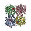 1h5rS S: Starting model for refinement |
|---|---|
| Similar structure data |


-Links
 PDBj
PDBj

- Assembly
| Deposited unit | 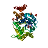
| ||||||||||||
|---|---|---|---|---|---|---|---|---|---|---|---|---|---|
| 1 |
| ||||||||||||
| 2 | 
| ||||||||||||
| Unit cell |
| ||||||||||||
| Components on special symmetry positions |
|
-Components
| #1: Protein | Mass: 32973.664 Da / Num. of mol.: 1 Source method: isolated from a genetically manipulated source Source: (gene. exp.) Aneurinibacillus thermoaerophilus (bacteria)Gene: rmlA / Plasmid: pTXB1 / Production host: References: UniProt: Q9AGY4, glucose-1-phosphate thymidylyltransferase | ||||
|---|---|---|---|---|---|
| #2: Chemical |   Type: DNA OH 5 prime terminus / Mass: 242.229 Da / Num. of mol.: 2 / Source method: obtained synthetically / Formula: C10H14N2O5 Type: DNA OH 5 prime terminus / Mass: 242.229 Da / Num. of mol.: 2 / Source method: obtained synthetically / Formula: C10H14N2O5#3: Chemical |   Mass: 96.063 Da / Num. of mol.: 3 / Source method: obtained synthetically / Formula: SO4 Mass: 96.063 Da / Num. of mol.: 3 / Source method: obtained synthetically / Formula: SO4#4: Water | ChemComp-HOH / |  Mass: 18.015 Da / Num. of mol.: 186 / Source method: isolated from a natural source / Formula: H2O Mass: 18.015 Da / Num. of mol.: 186 / Source method: isolated from a natural source / Formula: H2O |
-Experimental details
-Experiment
| Experiment | Method: X-RAY DIFFRACTION / Number of used crystals: 1 |
|---|
- Sample preparation
| Crystal | Density Matthews: 2.25 Å3/Da / Density % sol: 45.22 % |
|---|---|
| Crystal grow | Temperature: 293 K / Method: vapor diffusion, hanging drop / pH: 6.5 Details: 0.1M sodium cacodylate, 2.0M lithium sulfate, pH 6.5, VAPOR DIFFUSION, HANGING DROP, temperature 293K |
-Data collection
| Diffraction | Mean temperature: 110 K |
|---|---|
| Diffraction source | Source: SYNCHROTRON / Site: NSRRC  / Beamline: BL13C1 / Wavelength: 1 Å / Beamline: BL13C1 / Wavelength: 1 Å |
| Detector | Type: ADSC QUANTUM 315r / Detector: CCD / Date: Oct 9, 2011 / Details: mirrors |
| Radiation | Monochromator: Si / Protocol: SINGLE WAVELENGTH / Monochromatic (M) / Laue (L): M / Scattering type: x-ray |
| Radiation wavelength | Wavelength: 1 Å / Relative weight: 1 |
| Reflection | Resolution: 1.84→30 Å / Num. all: 26525 / Num. obs: 26483 / % possible obs: 99.5 % / Redundancy: 12.7 % / Rmerge(I) obs: 0.042 |
| Reflection shell | Resolution: 1.84→1.91 Å / Redundancy: 13 % / Rmerge(I) obs: 0.357 / % possible all: 100 |
- Processing
| Software |
| ||||||||||||||||||||||||||||||||||||
|---|---|---|---|---|---|---|---|---|---|---|---|---|---|---|---|---|---|---|---|---|---|---|---|---|---|---|---|---|---|---|---|---|---|---|---|---|---|
| Refinement | Method to determine structure: MOLECULAR REPLACEMENT Starting model: PDB entry 1H5R Resolution: 1.84→30 Å / Cor.coef. Fo:Fc: 0.956 / Cor.coef. Fo:Fc free: 0.935 / Occupancy max: 1 / Occupancy min: 0.5 / SU B: 2.919 / SU ML: 0.091 / Cross valid method: THROUGHOUT / σ(F): 0 / ESU R: 0.147 / ESU R Free: 0.137 / Stereochemistry target values: MAXIMUM LIKELIHOOD Details: HYDROGENS HAVE BEEN ADDED IN THE RIDING POSITIONS U VALUES: REFINED INDIVIDUALLY
| ||||||||||||||||||||||||||||||||||||
| Solvent computation | Ion probe radii: 0.8 Å / Shrinkage radii: 0.8 Å / VDW probe radii: 1.4 Å / Solvent model: MASK | ||||||||||||||||||||||||||||||||||||
| Displacement parameters | Biso max: 70.33 Å2 / Biso mean: 30.1519 Å2 / Biso min: 16.02 Å2
| ||||||||||||||||||||||||||||||||||||
| Refinement step | Cycle: LAST / Resolution: 1.84→30 Å
| ||||||||||||||||||||||||||||||||||||
| Refine LS restraints |
| ||||||||||||||||||||||||||||||||||||
| LS refinement shell | Resolution: 1.84→1.888 Å / Total num. of bins used: 20
|
