 Movie
Movie Controller

[English] 日本語
 Yorodumi
- PDB-4h4n: 1.1 Angstrom Crystal Structure of Hypothetical Protein BA_2335 fr... -
+ Open data

- Basic information
| Entry | Database: PDB / ID: 4h4n | ||||||
|---|---|---|---|---|---|---|---|
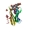
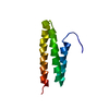
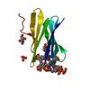
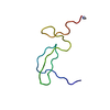
| Title | 1.1 Angstrom Crystal Structure of Hypothetical Protein BA_2335 from Bacillus anthracis | ||||||
 Components Components | hypothetical protein BA_2335 | ||||||
 Keywords Keywords | UNKNOWN FUNCTION / Structural Genomics / NIAID / National Institute of Allergy and Infectious Diseases / Center for Structural Genomics of Infectious Diseases / CSGID / hypothetical protein | ||||||
| Function / homology | Immunoglobulin-like - #3750 / : / BA_2335-like / Immunoglobulin-like / Sandwich / Mainly Beta / BETA-MERCAPTOETHANOL / Uncharacterized protein / Uncharacterized protein Function and homology information Function and homology information | ||||||
| Biological species |  | ||||||
| Method |  X-RAY DIFFRACTION / SYNCHROTRON / SAD / Resolution: 1.1 Å X-RAY DIFFRACTION / SYNCHROTRON / SAD / Resolution: 1.1 Å | ||||||
 Authors Authors | Minasov, G. / Wawrzak, Z. / Shuvalova, L. / Dubrovska, I. / Winsor, J. / Grimshaw, S. / Papazisi, L. / Anderson, W.F. / Center for Structural Genomics of Infectious Diseases (CSGID) | ||||||
 Citation Citation | Journal: TO BE PUBLISHED Title: 1.1 Angstrom Crystal Structure of Hypothetical Protein BA_2335 from Bacillus anthracis. Authors: Minasov, G. / Wawrzak, Z. / Shuvalova, L. / Dubrovska, I. / Winsor, J. / Grimshaw, S. / Papazisi, L. / Anderson, W.F. / Center for Structural Genomics of Infectious Diseases (CSGID) | ||||||
| History |
|
- Structure visualization
| Structure viewer | Molecule: MolmilJmol/JSmol |
|---|
- Downloads & links
-Download
| PDBx/mmCIF format | 4h4n.cif.gz | 44.1 KB | Display | PDBx/mmCIF format |
|---|---|---|---|---|
| PDB format | pdb4h4n.ent.gz | 31.2 KB | Display | PDB format |
| PDBx/mmJSON format | 4h4n.json.gz | Tree view | PDBx/mmJSON format | |
| Others |  Other downloads Other downloads |
-Validation report
| Arichive directory | https://data.pdbj.org/pub/pdb/validation_reports/h4/4h4nftp://data.pdbj.org/pub/pdb/validation_reports/h4/4h4n | HTTPS FTP |
|---|
-Related structure data
| Similar structure data | |
|---|---|
| Other databases |


-Links
 PDBj
PDBj


- Assembly
| Deposited unit | 
| ||||||||
|---|---|---|---|---|---|---|---|---|---|
| 1 |
| ||||||||
| Unit cell |
|
-Components
| #1: Protein | Mass: 7625.771 Da / Num. of mol.: 1 Source method: isolated from a genetically manipulated source Source: (gene. exp.) | ||||||||
|---|---|---|---|---|---|---|---|---|---|
| #2: Chemical |   Mass: 35.453 Da / Num. of mol.: 3 / Source method: obtained synthetically / Formula: Cl Mass: 35.453 Da / Num. of mol.: 3 / Source method: obtained synthetically / Formula: Cl#3: Chemical | ChemComp-BME / |   Mass: 78.133 Da / Num. of mol.: 1 / Source method: obtained synthetically / Formula: C2H6OS Mass: 78.133 Da / Num. of mol.: 1 / Source method: obtained synthetically / Formula: C2H6OS#4: Chemical | ChemComp-SO4 / |   Mass: 96.063 Da / Num. of mol.: 1 / Source method: obtained synthetically / Formula: SO4 Mass: 96.063 Da / Num. of mol.: 1 / Source method: obtained synthetically / Formula: SO4#5: Water | ChemComp-HOH / |  Mass: 18.015 Da / Num. of mol.: 87 / Source method: isolated from a natural source / Formula: H2O Mass: 18.015 Da / Num. of mol.: 87 / Source method: isolated from a natural source / Formula: H2OHas protein modification | N | |
-Experimental details
-Experiment
| Experiment | Method: X-RAY DIFFRACTION / Number of used crystals: 1 |
|---|
- Sample preparation
| Crystal | Density Matthews: 1.83 Å3/Da / Density % sol: 32.81 % |
|---|---|
| Crystal grow | Temperature: 295 K / Method: vapor diffusion, sitting drop / pH: 7.5 Details: Protein: 3.5mg/mL, 0.5M Sodium chloride, 0.01M Tris-HCl pH 8.3; Screen: Classics II (G4), 0.2M Lithium sulfate, 0.1M HEPES pH 7.5, 25% (w/v) PEG 3350., VAPOR DIFFUSION, SITTING DROP, temperature 295K |
-Data collection
| Diffraction | Mean temperature: 100 K |
|---|---|
| Diffraction source | Source: SYNCHROTRON / Site: APS  / Beamline: 21-ID-F / Wavelength: 0.97872 Å / Beamline: 21-ID-F / Wavelength: 0.97872 Å |
| Detector | Type: MARMOSAIC 225 mm CCD / Detector: CCD / Date: Aug 6, 2012 / Details: Beryllium lenses |
| Radiation | Monochromator: Diamond / Protocol: SINGLE WAVELENGTH / Monochromatic (M) / Laue (L): M / Scattering type: x-ray |
| Radiation wavelength | Wavelength: 0.97872 Å / Relative weight: 1 |
| Reflection | Resolution: 1.1→30 Å / Num. all: 23318 / Num. obs: 23318 / % possible obs: 99.1 % / Observed criterion σ(I): -3 / Redundancy: 5.2 % / Biso Wilson estimate: 10 Å2 / Rmerge(I) obs: 0.051 / Net I/σ(I): 26.3 |
| Reflection shell | Resolution: 1.1→1.12 Å / Redundancy: 3 % / Rmerge(I) obs: 0.333 / Mean I/σ(I) obs: 2.04 / Num. unique all: 1084 / % possible all: 93 |
- Processing
| Software |
| ||||||||||||||||||||||||||||||||||||||||||||||||||||||||||||||||||||||||||||||||||||||||||
|---|---|---|---|---|---|---|---|---|---|---|---|---|---|---|---|---|---|---|---|---|---|---|---|---|---|---|---|---|---|---|---|---|---|---|---|---|---|---|---|---|---|---|---|---|---|---|---|---|---|---|---|---|---|---|---|---|---|---|---|---|---|---|---|---|---|---|---|---|---|---|---|---|---|---|---|---|---|---|---|---|---|---|---|---|---|---|---|---|---|---|---|
| Refinement | Method to determine structure: SAD / Resolution: 1.1→26.01 Å / Cor.coef. Fo:Fc: 0.973 / Cor.coef. Fo:Fc free: 0.953 / SU B: 0.634 / SU ML: 0.014 Isotropic thermal model: Thermal Factors Anisotropically Refined Cross valid method: THROUGHOUT / ESU R: 0.033 / ESU R Free: 0.035 / Stereochemistry target values: MAXIMUM LIKELIHOOD / Details: HYDROGENS HAVE BEEN ADDED IN THE RIDING POSITIONS
| ||||||||||||||||||||||||||||||||||||||||||||||||||||||||||||||||||||||||||||||||||||||||||
| Solvent computation | Ion probe radii: 0.8 Å / Shrinkage radii: 0.8 Å / VDW probe radii: 1.2 Å / Solvent model: MASK | ||||||||||||||||||||||||||||||||||||||||||||||||||||||||||||||||||||||||||||||||||||||||||
| Displacement parameters | Biso mean: 11.142 Å2
| ||||||||||||||||||||||||||||||||||||||||||||||||||||||||||||||||||||||||||||||||||||||||||
| Refinement step | Cycle: LAST / Resolution: 1.1→26.01 Å
| ||||||||||||||||||||||||||||||||||||||||||||||||||||||||||||||||||||||||||||||||||||||||||
| Refine LS restraints |
| ||||||||||||||||||||||||||||||||||||||||||||||||||||||||||||||||||||||||||||||||||||||||||
| LS refinement shell | Resolution: 1.1→1.129 Å / Total num. of bins used: 20
|
