 Movie
Movie Controller

[English] 日本語
 Yorodumi
- PDB-4gv5: X-ray structure of crotamine, a cell-penetrating peptide from the... -
+ Open data

- Basic information
| Entry | Database: PDB / ID: 4gv5 | ||||||
|---|---|---|---|---|---|---|---|
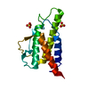
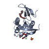
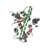
| Title | X-ray structure of crotamine, a cell-penetrating peptide from the Brazilian snake Crotalus durissus terrificus | ||||||
 Components Components | Crotamine Ile-19 | ||||||
 Keywords Keywords | TOXIN / alpha helix & beta sheet / Ion channel inhibitor / antibacterial peptide / Extracellular region | ||||||
| Function / homology |  Function and homology information Function and homology informationvenom-mediated inhibition of voltage-gated potassium channel activity / potassium channel regulator activity / defense response to fungus / toxin activity / killing of cells of another organism / defense response to bacterium / extracellular region Similarity search - Function | ||||||
| Biological species |  Crotalus durissus terrificus (tropical rattlesnake) Crotalus durissus terrificus (tropical rattlesnake) | ||||||
| Method |  X-RAY DIFFRACTION / SYNCHROTRON / SAD / Resolution: 1.7 Å X-RAY DIFFRACTION / SYNCHROTRON / SAD / Resolution: 1.7 Å | ||||||
 Authors Authors | Coronado, M.A. / Gabdulkhakov, A. / Georgieva, D. / Sankaran, B. / Murakami, M.T. / Arni, R.K. / Betzel, C. | ||||||
 Citation Citation | Journal: Acta Crystallogr.,Sect.D / Year: 2013 Title: Structure of the polypeptide crotamine from the Brazilian rattlesnake Crotalus durissus terrificus. Authors: Coronado, M.A. / Gabdulkhakov, A. / Georgieva, D. / Sankaran, B. / Murakami, M.T. / Arni, R.K. / Betzel, C. | ||||||
| History |
|
- Structure visualization
| Structure viewer | Molecule: MolmilJmol/JSmol |
|---|
- Downloads & links
-Download
| PDBx/mmCIF format | 4gv5.cif.gz | 68.9 KB | Display | PDBx/mmCIF format |
|---|---|---|---|---|
| PDB format | pdb4gv5.ent.gz | 53.2 KB | Display | PDB format |
| PDBx/mmJSON format | 4gv5.json.gz | Tree view | PDBx/mmJSON format | |
| Others |  Other downloads Other downloads |
-Validation report
| Arichive directory | https://data.pdbj.org/pub/pdb/validation_reports/gv/4gv5ftp://data.pdbj.org/pub/pdb/validation_reports/gv/4gv5 | HTTPS FTP |
|---|
-Related structure data
| Similar structure data |
|---|

-Links
 PDBj
PDBj
- Assembly
| Deposited unit | 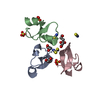
| |||||||||
|---|---|---|---|---|---|---|---|---|---|---|
| 1 |
| |||||||||
| 2 | 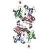
| |||||||||
| 3 | 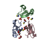
| |||||||||
| Unit cell |
| |||||||||
| Components on special symmetry positions |
|
-Components
| #1: Protein/peptide | Mass: 4902.878 Da / Num. of mol.: 3 / Source method: isolated from a natural source Source: (natural) Crotalus durissus terrificus (tropical rattlesnake)References: UniProt: P01475, UniProt: Q9PWF3*PLUS #2: Chemical | ChemComp-SCN /   Mass: 58.082 Da / Num. of mol.: 4 / Source method: obtained synthetically / Formula: CNS Mass: 58.082 Da / Num. of mol.: 4 / Source method: obtained synthetically / Formula: CNS#3: Chemical | ChemComp-GOL /   Mass: 92.094 Da / Num. of mol.: 4 / Source method: obtained synthetically / Formula: C3H8O3 Mass: 92.094 Da / Num. of mol.: 4 / Source method: obtained synthetically / Formula: C3H8O3#4: Chemical |   Mass: 96.063 Da / Num. of mol.: 3 / Source method: obtained synthetically / Formula: SO4 Mass: 96.063 Da / Num. of mol.: 3 / Source method: obtained synthetically / Formula: SO4#5: Water | ChemComp-HOH / |  Mass: 18.015 Da / Num. of mol.: 62 / Source method: isolated from a natural source / Formula: H2O Mass: 18.015 Da / Num. of mol.: 62 / Source method: isolated from a natural source / Formula: H2OHas protein modification | Y | |
|---|
-Experimental details
-Experiment
| Experiment | Method: X-RAY DIFFRACTION / Number of used crystals: 1 |
|---|
- Sample preparation
| Crystal | Density Matthews: 3.47 Å3/Da / Density % sol: 64.51 % |
|---|---|
| Crystal grow | Temperature: 293 K / Method: vapor diffusion, hanging drop / pH: 6.1 Details: 0.2 M sodium thiocyanate, 1.9 M ammonium sulphate, pH 6.1, VAPOR DIFFUSION, HANGING DROP, temperature 293.0K |
-Data collection
| Diffraction | Mean temperature: 100 K |
|---|---|
| Diffraction source | Source: SYNCHROTRON / Site: PETRA III, EMBL c/o DESY  / Beamline: P14 (MX2) / Wavelength: 1.12 / Beamline: P14 (MX2) / Wavelength: 1.12 |
| Detector | Type: RAYONIX MX225HE / Detector: CCD |
| Radiation | Protocol: SINGLE WAVELENGTH / Monochromatic (M) / Laue (L): M / Scattering type: x-ray |
| Radiation wavelength | Wavelength: 1.12 Å / Relative weight: 1 |
| Reflection | Resolution: 1.7→10 Å / Num. all: 22612 / Num. obs: 20890 / % possible obs: 97.7 % / Observed criterion σ(F): 2.5 / Observed criterion σ(I): 1 |
- Processing
| Software |
| ||||||||||||||||||||||||||||||||||||||||||||||||||||||||||||||||||||||||||||||||||||||||||||||||||||||||||||||||||||||||||||||||||||||||||||||||||||||||||||||||||||||||||
|---|---|---|---|---|---|---|---|---|---|---|---|---|---|---|---|---|---|---|---|---|---|---|---|---|---|---|---|---|---|---|---|---|---|---|---|---|---|---|---|---|---|---|---|---|---|---|---|---|---|---|---|---|---|---|---|---|---|---|---|---|---|---|---|---|---|---|---|---|---|---|---|---|---|---|---|---|---|---|---|---|---|---|---|---|---|---|---|---|---|---|---|---|---|---|---|---|---|---|---|---|---|---|---|---|---|---|---|---|---|---|---|---|---|---|---|---|---|---|---|---|---|---|---|---|---|---|---|---|---|---|---|---|---|---|---|---|---|---|---|---|---|---|---|---|---|---|---|---|---|---|---|---|---|---|---|---|---|---|---|---|---|---|---|---|---|---|---|---|---|---|---|
| Refinement | Method to determine structure: SAD / Resolution: 1.7→10 Å / Cor.coef. Fo:Fc: 0.975 / Cor.coef. Fo:Fc free: 0.958 / SU B: 4.493 / SU ML: 0.064 / Cross valid method: THROUGHOUT / ESU R: 0.096 / ESU R Free: 0.092 / Stereochemistry target values: MAXIMUM LIKELIHOOD / Details: HYDROGENS HAVE BEEN USED IF PRESENT IN THE INPUT
| ||||||||||||||||||||||||||||||||||||||||||||||||||||||||||||||||||||||||||||||||||||||||||||||||||||||||||||||||||||||||||||||||||||||||||||||||||||||||||||||||||||||||||
| Solvent computation | Ion probe radii: 0.8 Å / Shrinkage radii: 0.8 Å / VDW probe radii: 1.2 Å / Solvent model: MASK | ||||||||||||||||||||||||||||||||||||||||||||||||||||||||||||||||||||||||||||||||||||||||||||||||||||||||||||||||||||||||||||||||||||||||||||||||||||||||||||||||||||||||||
| Displacement parameters | Biso mean: 43.727 Å2
| ||||||||||||||||||||||||||||||||||||||||||||||||||||||||||||||||||||||||||||||||||||||||||||||||||||||||||||||||||||||||||||||||||||||||||||||||||||||||||||||||||||||||||
| Refinement step | Cycle: LAST / Resolution: 1.7→10 Å
| ||||||||||||||||||||||||||||||||||||||||||||||||||||||||||||||||||||||||||||||||||||||||||||||||||||||||||||||||||||||||||||||||||||||||||||||||||||||||||||||||||||||||||
| Refine LS restraints |
| ||||||||||||||||||||||||||||||||||||||||||||||||||||||||||||||||||||||||||||||||||||||||||||||||||||||||||||||||||||||||||||||||||||||||||||||||||||||||||||||||||||||||||
| LS refinement shell | Resolution: 1.7→1.743 Å / Total num. of bins used: 20
|
