 Movie
Movie Controller

[English] 日本語
 Yorodumi
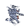
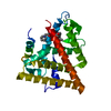
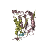
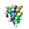
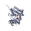
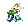
- PDB-4gkq: Structure of the neck and C-terminal motor homology domain of ViK... -
+ Open data

- Basic information
| Entry | Database: PDB / ID: 4gkq | ||||||
|---|---|---|---|---|---|---|---|
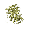
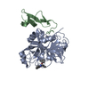
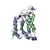
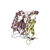
| Title | Structure of the neck and C-terminal motor homology domain of ViK1 from Candida glabrata | ||||||
 Components Components | SPINDLE POLE BODY-ASSOCIATED PROTEIN VIK1 | ||||||
 Keywords Keywords | STRUCTURAL PROTEIN / Kinesin motor domain-like fold / Microtubule binding protein / kinesin-associated protein / Kar3 | ||||||
| Function / homology |  Function and homology information Function and homology information | ||||||
| Biological species |  Candida glabrata (fungus) Candida glabrata (fungus) | ||||||
| Method |  X-RAY DIFFRACTION / SYNCHROTRON / MOLECULAR REPLACEMENT / Resolution: 2.99 Å X-RAY DIFFRACTION / SYNCHROTRON / MOLECULAR REPLACEMENT / Resolution: 2.99 Å | ||||||
 Authors Authors | Duan, D. / Allingham, J.S. | ||||||
 Citation Citation | Journal: J.Biol.Chem. / Year: 2012 Title: Neck Rotation and Neck Mimic Docking in the Noncatalytic Kar3-associated Protein Vik1. Authors: Duan, D. / Jia, Z. / Joshi, M. / Brunton, J. / Chan, M. / Drew, D. / Davis, D. / Allingham, J.S. | ||||||
| History |
|
- Structure visualization
| Structure viewer | Molecule: MolmilJmol/JSmol |
|---|
- Downloads & links
-Download
| PDBx/mmCIF format | 4gkq.cif.gz | 186.7 KB | Display | PDBx/mmCIF format |
|---|---|---|---|---|
| PDB format | pdb4gkq.ent.gz | 149.3 KB | Display | PDB format |
| PDBx/mmJSON format | 4gkq.json.gz | Tree view | PDBx/mmJSON format | |
| Others |  Other downloads Other downloads |
-Validation report
| Arichive directory | https://data.pdbj.org/pub/pdb/validation_reports/gk/4gkqftp://data.pdbj.org/pub/pdb/validation_reports/gk/4gkq | HTTPS FTP |
|---|
-Related structure data


-Links
 PDBj
PDBj- Assembly
| Deposited unit | 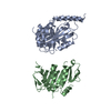
| ||||||||
|---|---|---|---|---|---|---|---|---|---|
| 1 | 
| ||||||||
| 2 | 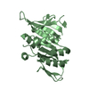
| ||||||||
| Unit cell |
|
-Components
| #1: Protein | Mass: 33260.988 Da / Num. of mol.: 2 / Fragment: UNP residues 302-584 Source method: isolated from a genetically manipulated source Source: (gene. exp.) Candida glabrata (fungus)Strain: ATCC 2001 / CBS 138 / JCM 3761 / NBRC 0622 / NRRL Y-65 Gene: CAGL0H00638g / Plasmid: pET16b / Production host:  #2: Water | ChemComp-HOH / |  Mass: 18.015 Da / Num. of mol.: 1 / Source method: isolated from a natural source / Formula: H2O Mass: 18.015 Da / Num. of mol.: 1 / Source method: isolated from a natural source / Formula: H2O |
|---|
-Experimental details
-Experiment
| Experiment | Method: X-RAY DIFFRACTION / Number of used crystals: 1 |
|---|
- Sample preparation
| Crystal | Density Matthews: 2.82 Å3/Da / Density % sol: 56.34 % |
|---|---|
| Crystal grow | Temperature: 277 K / Method: vapor diffusion, hanging drop / pH: 8.5 Details: 0.1M Tris, 12% PEG8000, 0.05M MgCl2, 0.15M NaCl, 5% Ethylene glycol, 1mM TCEP, pH 8.5, VAPOR DIFFUSION, HANGING DROP, temperature 277K |
-Data collection
| Diffraction | Mean temperature: 100 K | |||||||||||||||||||||||||||||||||||||||||||||||||||||||||||||||||||||||||||||||||||||||||||||||||||||||||||||||||||||||||||||||||||||||||||||||||||
|---|---|---|---|---|---|---|---|---|---|---|---|---|---|---|---|---|---|---|---|---|---|---|---|---|---|---|---|---|---|---|---|---|---|---|---|---|---|---|---|---|---|---|---|---|---|---|---|---|---|---|---|---|---|---|---|---|---|---|---|---|---|---|---|---|---|---|---|---|---|---|---|---|---|---|---|---|---|---|---|---|---|---|---|---|---|---|---|---|---|---|---|---|---|---|---|---|---|---|---|---|---|---|---|---|---|---|---|---|---|---|---|---|---|---|---|---|---|---|---|---|---|---|---|---|---|---|---|---|---|---|---|---|---|---|---|---|---|---|---|---|---|---|---|---|---|---|---|---|
| Diffraction source | Source: SYNCHROTRON / Site: APS  / Beamline: 23-ID-B / Wavelength: 1.033 Å / Beamline: 23-ID-B / Wavelength: 1.033 Å | |||||||||||||||||||||||||||||||||||||||||||||||||||||||||||||||||||||||||||||||||||||||||||||||||||||||||||||||||||||||||||||||||||||||||||||||||||
| Detector | Type: MARMOSAIC 300 mm CCD / Detector: CCD / Date: Jul 15, 2010 | |||||||||||||||||||||||||||||||||||||||||||||||||||||||||||||||||||||||||||||||||||||||||||||||||||||||||||||||||||||||||||||||||||||||||||||||||||
| Radiation | Monochromator: Double crystal cryo-cooled Si (111) / Protocol: SINGLE WAVELENGTH / Monochromatic (M) / Laue (L): M / Scattering type: x-ray | |||||||||||||||||||||||||||||||||||||||||||||||||||||||||||||||||||||||||||||||||||||||||||||||||||||||||||||||||||||||||||||||||||||||||||||||||||
| Radiation wavelength | Wavelength: 1.033 Å / Relative weight: 1 | |||||||||||||||||||||||||||||||||||||||||||||||||||||||||||||||||||||||||||||||||||||||||||||||||||||||||||||||||||||||||||||||||||||||||||||||||||
| Reflection | Resolution: 2.99→20 Å / Num. all: 14671 / Num. obs: 14501 / % possible obs: 98.8 % / Observed criterion σ(F): 0 / Observed criterion σ(I): -3 / Redundancy: 5.8 % / Biso Wilson estimate: 103.571 Å2 / Rmerge(I) obs: 0.088 / Net I/σ(I): 13.14 | |||||||||||||||||||||||||||||||||||||||||||||||||||||||||||||||||||||||||||||||||||||||||||||||||||||||||||||||||||||||||||||||||||||||||||||||||||
| Reflection shell | Diffraction-ID: 1
|
- Processing
| Software |
| |||||||||||||||||||||||||||||||||||||||||||||||||||||||||||||||||||||||||||
|---|---|---|---|---|---|---|---|---|---|---|---|---|---|---|---|---|---|---|---|---|---|---|---|---|---|---|---|---|---|---|---|---|---|---|---|---|---|---|---|---|---|---|---|---|---|---|---|---|---|---|---|---|---|---|---|---|---|---|---|---|---|---|---|---|---|---|---|---|---|---|---|---|---|---|---|---|
| Refinement | Method to determine structure: MOLECULAR REPLACEMENT / Resolution: 2.99→19.8 Å / Cor.coef. Fo:Fc: 0.94 / Cor.coef. Fo:Fc free: 0.921 / WRfactor Rfree: 0.2896 / WRfactor Rwork: 0.2354 / Occupancy max: 1 / Occupancy min: 1 / FOM work R set: 0.806 / SU B: 39.247 / SU ML: 0.33 / SU R Cruickshank DPI: 0.3711 / SU Rfree: 0.4068 / Cross valid method: THROUGHOUT / σ(F): 0 / ESU R Free: 0.407 / Stereochemistry target values: MAXIMUM LIKELIHOOD / Details: HYDROGENS HAVE BEEN USED IF PRESENT IN THE INPUT
| |||||||||||||||||||||||||||||||||||||||||||||||||||||||||||||||||||||||||||
| Solvent computation | Ion probe radii: 0.8 Å / Shrinkage radii: 0.8 Å / VDW probe radii: 1.2 Å / Solvent model: MASK | |||||||||||||||||||||||||||||||||||||||||||||||||||||||||||||||||||||||||||
| Displacement parameters | Biso max: 201.29 Å2 / Biso mean: 113.981 Å2 / Biso min: 66.06 Å2
| |||||||||||||||||||||||||||||||||||||||||||||||||||||||||||||||||||||||||||
| Refinement step | Cycle: LAST / Resolution: 2.99→19.8 Å
| |||||||||||||||||||||||||||||||||||||||||||||||||||||||||||||||||||||||||||
| Refine LS restraints |
| |||||||||||||||||||||||||||||||||||||||||||||||||||||||||||||||||||||||||||
| LS refinement shell | Resolution: 2.99→3.066 Å / Total num. of bins used: 20
| |||||||||||||||||||||||||||||||||||||||||||||||||||||||||||||||||||||||||||
| Refinement TLS params. | Method: refined / Refine-ID: X-RAY DIFFRACTION
| |||||||||||||||||||||||||||||||||||||||||||||||||||||||||||||||||||||||||||
| Refinement TLS group |
|
