 Movie
Movie Controller


+ Open data

- Basic information
| Entry | Database: PDB / ID: 4fs6 | ||||||||||||||||||
|---|---|---|---|---|---|---|---|---|---|---|---|---|---|---|---|---|---|---|---|
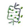
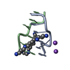
| Title | Crystal structure of the Z-DNA hexamer CGCGCG at 500 mM CaCl2 | ||||||||||||||||||
 Components Components | DNA (5'-D(* Keywords KeywordsDNA / Z-DNA / Ca2+ binding | Function / homology | DNA |  Function and homology information Function and homology informationMethod |  X-RAY DIFFRACTION / SYNCHROTRON / MOLECULAR REPLACEMENT / Resolution: 1.3 Å X-RAY DIFFRACTION / SYNCHROTRON / MOLECULAR REPLACEMENT / Resolution: 1.3 Å  Authors AuthorsChatake, T. / Sunami, T. |  CitationJournal: J.Inorg.Biochem. / Year: 2013 CitationJournal: J.Inorg.Biochem. / Year: 2013Title: Direct interactions between Z-DNA and alkaline earth cations, discovered in the presence of high concentrations of MgCl2 and CaCl2 Authors: Chatake, T. / Sunami, T. History |
|
- Structure visualization
| Structure viewer | Molecule: MolmilJmol/JSmol |
|---|
- Downloads & links
-Download
| PDBx/mmCIF format | 4fs6.cif.gz | 17.8 KB | Display | PDBx/mmCIF format |
|---|---|---|---|---|
| PDB format | pdb4fs6.ent.gz | 11.2 KB | Display | PDB format |
| PDBx/mmJSON format | 4fs6.json.gz | Tree view | PDBx/mmJSON format | |
| Others |  Other downloads Other downloads |
-Validation report
| Arichive directory | https://data.pdbj.org/pub/pdb/validation_reports/fs/4fs6ftp://data.pdbj.org/pub/pdb/validation_reports/fs/4fs6 | HTTPS FTP |
|---|
-Related structure data


-Links
 PDBj
PDBj

- Assembly
| Deposited unit | 
| |||||||||
|---|---|---|---|---|---|---|---|---|---|---|
| 1 | 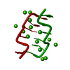
| |||||||||
| Unit cell |
| |||||||||
| Components on special symmetry positions |
|
-Components
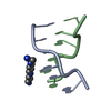
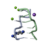
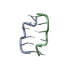
| #1: DNA chain | Mass: 1810.205 Da / Num. of mol.: 1 / Source method: obtained synthetically / Details: Chemical synthesis | ||||
|---|---|---|---|---|---|
| #2: Chemical | ChemComp-CA /   Mass: 40.078 Da / Num. of mol.: 5 / Source method: obtained synthetically / Formula: Ca Mass: 40.078 Da / Num. of mol.: 5 / Source method: obtained synthetically / Formula: Ca#3: Chemical |   Mass: 35.453 Da / Num. of mol.: 2 / Source method: obtained synthetically / Formula: Cl Mass: 35.453 Da / Num. of mol.: 2 / Source method: obtained synthetically / Formula: Cl#4: Water | ChemComp-HOH / |  Mass: 18.015 Da / Num. of mol.: 41 / Source method: isolated from a natural source / Formula: H2O Mass: 18.015 Da / Num. of mol.: 41 / Source method: isolated from a natural source / Formula: H2O |
-Experimental details
-Experiment
| Experiment | Method: X-RAY DIFFRACTION / Number of used crystals: 1 |
|---|
- Sample preparation
| Crystal | Density Matthews: 1.94 Å3/Da / Density % sol: 36.66 % |
|---|---|
| Crystal grow | Temperature: 293 K / Method: temperature control / pH: 7 Details: 2.0mM DNA, 20mM Na cacodylate, 30% 2-methyl-2,4-pentanediol, 500mM CaCl2, pH 7.0, TEMPERATURE CONTROL, temperature 293K |
-Data collection
| Diffraction | Mean temperature: 100 K |
|---|---|
| Diffraction source | Source: SYNCHROTRON / Site: SPring-8  / Beamline: BL38B1 / Wavelength: 1 Å / Beamline: BL38B1 / Wavelength: 1 Å |
| Detector | Type: ADSC QUANTUM 315 / Detector: CCD / Date: Feb 13, 2008 |
| Radiation | Monochromator: Fixed exit Si (111) double crystal monochromator Protocol: SINGLE WAVELENGTH / Monochromatic (M) / Laue (L): M / Scattering type: x-ray |
| Radiation wavelength | Wavelength: 1 Å / Relative weight: 1 |
| Reflection twin | Operator: -h,-k,l / Fraction: 0.31 |
| Reflection | Resolution: 1.3→36 Å / Num. all: 3879 / Num. obs: 3859 / % possible obs: 99.2 % / Observed criterion σ(F): 0 / Observed criterion σ(I): 0 / Redundancy: 6.6 % / Biso Wilson estimate: 7.49 Å2 / Rmerge(I) obs: 0.072 |
| Reflection shell | Resolution: 1.3→1.47 Å / Redundancy: 8.2 % / Rmerge(I) obs: 0.051 / Num. unique all: 539 / % possible all: 99.7 |
- Processing
| Software |
| ||||||||||||||||||||||||
|---|---|---|---|---|---|---|---|---|---|---|---|---|---|---|---|---|---|---|---|---|---|---|---|---|---|
| Refinement | Method to determine structure: MOLECULAR REPLACEMENT / Resolution: 1.3→23.821 Å / Occupancy max: 1 / Occupancy min: 0.25 / FOM work R set: 0.7962 / σ(F): 2.92 / Phase error: 25.91 / Stereochemistry target values: TWIN_LSQ_F
| ||||||||||||||||||||||||
| Solvent computation | Shrinkage radii: 0 Å / VDW probe radii: 0.3 Å / Solvent model: FLAT BULK SOLVENT MODEL / Bsol: 71.397 Å2 / ksol: 0.6 e/Å3 | ||||||||||||||||||||||||
| Displacement parameters | Biso max: 80.37 Å2 / Biso mean: 12.72 Å2 / Biso min: 1.26 Å2
| ||||||||||||||||||||||||
| Refinement step | Cycle: LAST / Resolution: 1.3→23.821 Å
| ||||||||||||||||||||||||
| Refine LS restraints |
| ||||||||||||||||||||||||
| LS refinement shell | Resolution: 1.3002→13.2757 Å / Total num. of bins used: 1
|
