[English] 日本語
Yorodumi
- PDB-4fs1: Base pairing mechanism of N2,3-ethenoguanine with dTTP by human p... -

+
Open data


ID or keywords:

Loading...

-
Basic information

Entry
Database: PDB / ID: 4fs1
Title
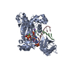
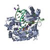
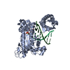
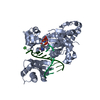
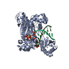
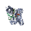
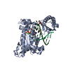
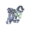
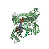
Base pairing mechanism of N2,3-ethenoguanine with dTTP by human polymerase iota
Components
  • DNA 5'-D(*TP*CP*TP*(EFG)P*GP*GP*GP*TP*CP*CP*TP*AP*GP*GP*AP*CP*CP*(DOC))-3'
  • DNA polymerase iotaPOLI
KeywordsTRANSFERASE/DNA / human DNA polymerase iota / DNA polymerase / DNA replication / lesion bypass / 2'-fluoro arabinose / N2 / 3-ethenoguanine / dTTP / template / TRANSFERASE-DNA complex
Function / homology
Function and homology information


translesion synthesis / error-prone translesion synthesis / Translesion synthesis by POLI / Termination of translesion DNA synthesis / cytoplasmic ribonucleoprotein granule / DNA replication / damaged DNA binding / DNA-directed DNA polymerase / DNA-directed DNA polymerase activity / nuclear speck ...translesion synthesis / error-prone translesion synthesis / Translesion synthesis by POLI / Termination of translesion DNA synthesis / cytoplasmic ribonucleoprotein granule / DNA replication / damaged DNA binding / DNA-directed DNA polymerase / DNA-directed DNA polymerase activity / nuclear speck / DNA repair / nucleoplasm / metal ion binding
Similarity search - Function
DNA polymerase, Y-family, little finger domain / MutS, DNA mismatch repair protein, domain I - #60 / MutS, DNA mismatch repair protein, domain I / DNA polymerase, Y-family, little finger domain / impB/mucB/samB family C-terminal domain / UmuC domain / DNA polymerase, Y-family, little finger domain superfamily / impB/mucB/samB family / UmuC domain profile. / Reverse transcriptase/Diguanylate cyclase domain ...DNA polymerase, Y-family, little finger domain / MutS, DNA mismatch repair protein, domain I - #60 / MutS, DNA mismatch repair protein, domain I / DNA polymerase, Y-family, little finger domain / impB/mucB/samB family C-terminal domain / UmuC domain / DNA polymerase, Y-family, little finger domain superfamily / impB/mucB/samB family / UmuC domain profile. / Reverse transcriptase/Diguanylate cyclase domain / Dna Ligase; domain 1 / 5' to 3' exonuclease, C-terminal subdomain / DNA polymerase; domain 1 / Reverse transcriptase/Diguanylate cyclase domain / Alpha-Beta Plaits / DNA/RNA polymerase superfamily / 2-Layer Sandwich / Orthogonal Bundle / 3-Layer(aba) Sandwich / Mainly Alpha / Alpha Beta
Similarity search - Domain/homology
THYMIDINE-5'-TRIPHOSPHATE / DNA / DNA (> 10) / DNA polymerase iota
Similarity search - Component
Biological speciesHomo sapiens (human)
MethodX-RAY DIFFRACTION / SYNCHROTRON / MOLECULAR REPLACEMENT / Resolution: 2.5 Å
AuthorsZhao, L.
CitationJournal: J.Biol.Chem. / Year: 2012
Title: Basis of Miscoding of the DNA Adduct N2,3-Ethenoguanine by Human Y-family DNA Polymerases.
Authors: Zhao, L. / Pence, M.G. / Christov, P.P. / Wawrzak, Z. / Choi, J.Y. / Rizzo, C.J. / Egli, M. / Guengerich, F.P.
History
DepositionJun 26, 2012Deposition site: RCSB / Processing site: RCSB
Revision 1.0Aug 29, 2012Provider: repository / Type: Initial release
Revision 1.1Sep 12, 2012Group: Database references
Revision 1.2Oct 31, 2012Group: Database references
Revision 1.3Feb 28, 2024Group: Data collection / Database references / Derived calculations
Category: chem_comp_atom / chem_comp_bond ...chem_comp_atom / chem_comp_bond / database_2 / pdbx_struct_conn_angle / struct_conn / struct_site
Item: _database_2.pdbx_DOI / _database_2.pdbx_database_accession ..._database_2.pdbx_DOI / _database_2.pdbx_database_accession / _pdbx_struct_conn_angle.ptnr1_auth_asym_id / _pdbx_struct_conn_angle.ptnr1_auth_comp_id / _pdbx_struct_conn_angle.ptnr1_auth_seq_id / _pdbx_struct_conn_angle.ptnr1_label_asym_id / _pdbx_struct_conn_angle.ptnr1_label_atom_id / _pdbx_struct_conn_angle.ptnr1_label_comp_id / _pdbx_struct_conn_angle.ptnr1_label_seq_id / _pdbx_struct_conn_angle.ptnr2_auth_seq_id / _pdbx_struct_conn_angle.ptnr2_label_asym_id / _pdbx_struct_conn_angle.ptnr3_auth_asym_id / _pdbx_struct_conn_angle.ptnr3_auth_comp_id / _pdbx_struct_conn_angle.ptnr3_auth_seq_id / _pdbx_struct_conn_angle.ptnr3_label_asym_id / _pdbx_struct_conn_angle.ptnr3_label_atom_id / _pdbx_struct_conn_angle.ptnr3_label_comp_id / _pdbx_struct_conn_angle.ptnr3_label_seq_id / _pdbx_struct_conn_angle.value / _struct_conn.pdbx_dist_value / _struct_conn.pdbx_leaving_atom_flag / _struct_conn.ptnr1_auth_asym_id / _struct_conn.ptnr1_auth_comp_id / _struct_conn.ptnr1_auth_seq_id / _struct_conn.ptnr1_label_asym_id / _struct_conn.ptnr1_label_atom_id / _struct_conn.ptnr1_label_comp_id / _struct_conn.ptnr1_label_seq_id / _struct_conn.ptnr2_auth_comp_id / _struct_conn.ptnr2_auth_seq_id / _struct_conn.ptnr2_label_asym_id / _struct_conn.ptnr2_label_atom_id / _struct_conn.ptnr2_label_comp_id / _struct_site.pdbx_auth_asym_id / _struct_site.pdbx_auth_comp_id / _struct_site.pdbx_auth_seq_id

-
Structure visualization

Structure viewerMolecule:
MolmilJmol/JSmol

Downloads & links

-
Assembly

Deposited unit
B: DNA 5'-D(*TP*CP*TP*(EFG)P*GP*GP*GP*TP*CP*CP*TP*AP*GP*GP*AP*CP*CP*(DOC))-3'
C: DNA 5'-D(*TP*CP*TP*(EFG)P*GP*GP*GP*TP*CP*CP*TP*AP*GP*GP*AP*CP*CP*(DOC))-3'
A: DNA polymerase iota
hetero molecules


Theoretical massNumber of molelcules
Total (without water)58,6758
Polymers58,0963
Non-polymers5785
Water1,17165
1


  • Idetical with deposited unit
  • defined by author&software
TypeNameSymmetry operationNumber
identity operation1_555x,y,z1
Buried area5160 Å2
ΔGint-66 kcal/mol
Surface area20020 Å2
MethodPISA
Unit cell
Length a, b, c (Å)97.470, 97.470, 203.541
Angle α, β, γ (deg.)90.00, 90.00, 120.00
Int Tables number179
Space group name H-MP6522

-
Components

-
DNA chain / Protein , 2 types, 3 molecules BCA

#1: DNA chain DNA 5'-D(*TP*CP*TP*(EFG)P*GP*GP*GP*TP*CP*CP*TP*AP*GP*GP*AP*CP*CP*(DOC))-3'


Mass: 5534.566 Da / Num. of mol.: 2 / Source method: obtained synthetically / Details: site-specifically modified oligonucleotide
#2: Protein DNA polymerase iota / POLI / Eta2 / RAD30 homolog B


Mass: 47027.344 Da / Num. of mol.: 1
Source method: isolated from a genetically manipulated source
Source: (gene. exp.) Homo sapiens (human) / Gene: POLI, RAD30B / Production host: Escherichia coli (E. coli) / Strain (production host): BL21 GOLD (DE3) / References: UniProt: Q9UNA4, DNA-directed DNA polymerase

-
Non-polymers , 4 types, 70 molecules

#3: Chemical ChemComp-MG / MAGNESIUM ION


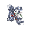
Mass: 24.305 Da / Num. of mol.: 3 / Source method: obtained synthetically / Formula: Mg
#4: Chemical ChemComp-NA / SODIUM ION


Mass: 22.990 Da / Num. of mol.: 1 / Source method: obtained synthetically / Formula: Na
#5: Chemical ChemComp-TTP / THYMIDINE-5'-TRIPHOSPHATE / Thymidine triphosphate


Mass: 482.168 Da / Num. of mol.: 1 / Source method: obtained synthetically / Formula: C10H17N2O14P3
#6: Water ChemComp-HOH / water / Water


Mass: 18.015 Da / Num. of mol.: 65 / Source method: isolated from a natural source / Formula: H2O

-
Experimental details

-
Experiment

ExperimentMethod: X-RAY DIFFRACTION / Number of used crystals: 1

-
Sample preparation

CrystalDensity Matthews: 2.4 Å3/Da / Density % sol: 48.79 %
Crystal growTemperature: 277 K / pH: 6.9
Details: 210 microM polymerase iota, 253 microM DNA complex, 10 mM MgCl2, 20 mM dTTP; mix 1:1 ratio with precipitant, 0.1 M MES (pH 6.5), 0.3 M (NH4)2SO4, 17% PEG 5000, VAPOR DIFFUSION, HANGING DROP, temperature 277K

-
Data collection

DiffractionMean temperature: 110 K
Diffraction sourceSource: SYNCHROTRON / Site: APS / Beamline: 21-ID-G / Wavelength: 0.97857
DetectorType: MAR scanner 300 mm plate / Detector: IMAGE PLATE / Date: Jun 9, 2012
RadiationMonochromator: C(111) / Protocol: SINGLE WAVELENGTH / Monochromatic (M) / Laue (L): M / Scattering type: x-ray
Radiation wavelengthWavelength: 0.97857 Å / Relative weight: 1
ReflectionResolution: 2.5→84.41 Å / Num. obs: 20822 / % possible obs: 99.8 % / Observed criterion σ(I): 2.75
Reflection shellResolution: 2.49→2.53 Å / % possible all: 100

-
Processing

Software
NameVersionClassification
EMBL(with LS-CAT developed extensions)data collection
MOLREP11.0.02phasing
REFMAC5.6.0117refinement
HKL-2000data reduction
HKL-2000data scaling
RefinementMethod to determine structure: MOLECULAR REPLACEMENT / Resolution: 2.5→84.41 Å / Cor.coef. Fo:Fc: 0.944 / Cor.coef. Fo:Fc free: 0.92 / SU B: 17.877 / SU ML: 0.214 / Cross valid method: THROUGHOUT / ESU R: 0.413 / ESU R Free: 0.287 / Stereochemistry target values: MAXIMUM LIKELIHOOD
RfactorNum. reflection% reflectionSelection details
Rfree0.27 1063 5.2 %RANDOM
Rwork0.215 ---
obs0.218 19448 99.7 %-
all-20575 --
Solvent computationIon probe radii: 0.8 Å / Shrinkage radii: 0.8 Å / VDW probe radii: 1.2 Å / Solvent model: MASK
Displacement parametersBiso mean: 56.66 Å2
Baniso -1Baniso -2Baniso -3
1-1.37 Å20.68 Å20 Å2
2--1.37 Å20 Å2
3----2.05 Å2
Refinement stepCycle: LAST / Resolution: 2.5→84.41 Å
ProteinNucleic acidLigandSolventTotal
Num. atoms2944 328 33 65 3370
Refine LS restraints
Refine-IDTypeDev idealDev ideal targetNumber
X-RAY DIFFRACTIONr_bond_refined_d0.0140.0193381
X-RAY DIFFRACTIONr_bond_other_d
X-RAY DIFFRACTIONr_angle_refined_deg1.9911.9484642
X-RAY DIFFRACTIONr_angle_other_deg
X-RAY DIFFRACTIONr_dihedral_angle_1_deg6.615381
X-RAY DIFFRACTIONr_dihedral_angle_2_deg40.40424.839124
X-RAY DIFFRACTIONr_dihedral_angle_3_deg20.41315553
X-RAY DIFFRACTIONr_dihedral_angle_4_deg24.8391519
X-RAY DIFFRACTIONr_chiral_restr0.1390.2534
X-RAY DIFFRACTIONr_gen_planes_refined0.0080.0212363
X-RAY DIFFRACTIONr_gen_planes_other
X-RAY DIFFRACTIONr_nbd_refined
X-RAY DIFFRACTIONr_nbd_other
X-RAY DIFFRACTIONr_nbtor_refined
X-RAY DIFFRACTIONr_nbtor_other
X-RAY DIFFRACTIONr_xyhbond_nbd_refined
X-RAY DIFFRACTIONr_xyhbond_nbd_other
X-RAY DIFFRACTIONr_metal_ion_refined
X-RAY DIFFRACTIONr_metal_ion_other
X-RAY DIFFRACTIONr_symmetry_vdw_refined
X-RAY DIFFRACTIONr_symmetry_vdw_other
X-RAY DIFFRACTIONr_symmetry_hbond_refined
X-RAY DIFFRACTIONr_symmetry_hbond_other
X-RAY DIFFRACTIONr_symmetry_metal_ion_refined
X-RAY DIFFRACTIONr_symmetry_metal_ion_other
X-RAY DIFFRACTIONr_mcbond_it
X-RAY DIFFRACTIONr_mcbond_other
X-RAY DIFFRACTIONr_mcangle_it
X-RAY DIFFRACTIONr_scbond_it
X-RAY DIFFRACTIONr_scangle_it
X-RAY DIFFRACTIONr_rigid_bond_restr
X-RAY DIFFRACTIONr_sphericity_free
X-RAY DIFFRACTIONr_sphericity_bonded
LS refinement shellResolution: 2.5→2.56 Å / Total num. of bins used: 20
RfactorNum. reflection% reflection
Rfree0.374 73 -
Rwork0.311 1224 -
obs--100 %
Refinement TLS params.

Method: refined / Refine-ID: X-RAY DIFFRACTION

IDL112)L122)L132)L222)L232)L332)S11 (Å °)S12 (Å °)S13 (Å °)S21 (Å °)S22 (Å °)S23 (Å °)S31 (Å °)S32 (Å °)S33 (Å °)T112)T122)T132)T222)T232)T332)Origin x (Å)Origin y (Å)Origin z (Å)
10.7077-0.7323-0.95242.95721.512.0233-0.07850.2560.16690.27480.0093-0.35580.1387-0.29490.06930.09430.0289-0.03430.1450.03670.235-4.72536.1462-19.6792
20.2164-0.09560.05480.7554-0.08930.4480.03730.04870.0438-0.0807-0.0739-0.15940.0639-0.06230.03660.06930.03880.01030.07150.01180.0391-7.288526.9974-15.2554
Refinement TLS group
IDRefine-IDRefine TLS-IDAuth asym-IDAuth seq-ID
1X-RAY DIFFRACTION1C839 - 847
2X-RAY DIFFRACTION2A26 - 414

+
About Yorodumi

-
News

-
Feb 9, 2022. New format data for meta-information of EMDB entries

New format data for meta-information of EMDB entries

  • Version 3 of the EMDB header file is now the official format.
  • The previous official version 1.9 will be removed from the archive.

Related info.:EMDB header

External links:wwPDB to switch to version 3 of the EMDB data model

-
Aug 12, 2020. Covid-19 info

Covid-19 info

URL: https://pdbj.org/emnavi/covid19.php

New page: Covid-19 featured information page in EM Navigator.

Related info.:Covid-19 info / Mar 5, 2020. Novel coronavirus structure data

+
Mar 5, 2020. Novel coronavirus structure data

Novel coronavirus structure data

Related info.:Yorodumi Speices / Aug 12, 2020. Covid-19 info

External links:COVID-19 featured content - PDBj / Molecule of the Month (242):Coronavirus Proteases

+
Jan 31, 2019. EMDB accession codes are about to change! (news from PDBe EMDB page)

EMDB accession codes are about to change! (news from PDBe EMDB page)

  • The allocation of 4 digits for EMDB accession codes will soon come to an end. Whilst these codes will remain in use, new EMDB accession codes will include an additional digit and will expand incrementally as the available range of codes is exhausted. The current 4-digit format prefixed with “EMD-” (i.e. EMD-XXXX) will advance to a 5-digit format (i.e. EMD-XXXXX), and so on. It is currently estimated that the 4-digit codes will be depleted around Spring 2019, at which point the 5-digit format will come into force.
  • The EM Navigator/Yorodumi systems omit the EMD- prefix.

Related info.:Q: What is EMD? / ID/Accession-code notation in Yorodumi/EM Navigator

External links:EMDB Accession Codes are Changing Soon! / Contact to PDBj

+
Jul 12, 2017. Major update of PDB

Major update of PDB

  • wwPDB released updated PDB data conforming to the new PDBx/mmCIF dictionary.
  • This is a major update changing the version number from 4 to 5, and with Remediation, in which all the entries are updated.
  • In this update, many items about electron microscopy experimental information are reorganized (e.g. em_software).
  • Now, EM Navigator and Yorodumi are based on the updated data.

External links:wwPDB Remediation / Enriched Model Files Conforming to OneDep Data Standards Now Available in the PDB FTP Archive

-
Yorodumi

Thousand views of thousand structures

  • Yorodumi is a browser for structure data from EMDB, PDB, SASBDB, etc.
  • This page is also the successor to EM Navigator detail page, and also detail information page/front-end page for Omokage search.
  • The word "yorodu" (or yorozu) is an old Japanese word meaning "ten thousand". "mi" (miru) is to see.

Related info.:EMDB / PDB / SASBDB / Comparison of 3 databanks / Yorodumi Search / Aug 31, 2016. New EM Navigator & Yorodumi / Yorodumi Papers / Jmol/JSmol / Function and homology information / Changes in new EM Navigator and Yorodumi

Read more