 Movie
Movie Controller


+ Open data

- Basic information
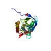
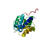
| Entry | Database: PDB / ID: 4fqa | ||||||
|---|---|---|---|---|---|---|---|
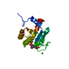
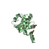
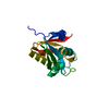
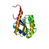
| Title | Crystal structure of toxic effector Tse1 | ||||||
 Components Components | toxic effector Tse1 | ||||||
 Keywords Keywords | TOXIN / helix and strand / toxic effector / Tsi1 | ||||||
| Function / homology |  Function and homology information Function and homology informationgamma-D-glutamyl-meso-diaminopimelate peptidase / amidase activity / host cell membrane / extracellular region / membrane Similarity search - Function | ||||||
| Biological species |  Pseudomonas aeruginosa PAO1 (bacteria) Pseudomonas aeruginosa PAO1 (bacteria) | ||||||
| Method |  X-RAY DIFFRACTION / SIRAS / Resolution: 2.1 Å X-RAY DIFFRACTION / SIRAS / Resolution: 2.1 Å | ||||||
 Authors Authors | Li, L. / Zhang, W. / Wang, T. | ||||||
 Citation Citation | Journal: To be Published Title: Structural insight on toxic effector Tse1 Authors: Li, L. / Zhang, W. / Wang, T. | ||||||
| History |
|
- Structure visualization
| Structure viewer | Molecule: MolmilJmol/JSmol |
|---|
- Downloads & links
-Download
| PDBx/mmCIF format | 4fqa.cif.gz | 72.5 KB | Display | PDBx/mmCIF format |
|---|---|---|---|---|
| PDB format | pdb4fqa.ent.gz | 54 KB | Display | PDB format |
| PDBx/mmJSON format | 4fqa.json.gz | Tree view | PDBx/mmJSON format | |
| Others |  Other downloads Other downloads |
-Validation report
| Arichive directory | https://data.pdbj.org/pub/pdb/validation_reports/fq/4fqaftp://data.pdbj.org/pub/pdb/validation_reports/fq/4fqa | HTTPS FTP |
|---|
-Related structure data
| Related structure data | |
|---|---|
| Similar structure data |


-Links
 PDBj
PDBj- Assembly
| Deposited unit | 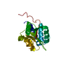
| ||||||||
|---|---|---|---|---|---|---|---|---|---|
| 1 |
| ||||||||
| Unit cell |
| ||||||||
| Details | monomer binds with Tsi1 |
-Components
| #1: Protein | Mass: 17529.828 Da / Num. of mol.: 1 Source method: isolated from a genetically manipulated source Details: 6His-tagged / Source: (gene. exp.) Pseudomonas aeruginosa PAO1 (bacteria) / Gene: PA1844 / Plasmid: pET29b(+) / Production host: |
|---|---|
| #2: Water | ChemComp-HOH /  Mass: 18.015 Da / Num. of mol.: 120 / Source method: isolated from a natural source / Formula: H2O Mass: 18.015 Da / Num. of mol.: 120 / Source method: isolated from a natural source / Formula: H2O |
| Has protein modification | Y |
-Experimental details
-Experiment
| Experiment | Method: X-RAY DIFFRACTION / Number of used crystals: 2 |
|---|
- Sample preparation
| Crystal | Density Matthews: 2.12 Å3/Da / Density % sol: 42.01 % |
|---|---|
| Crystal grow | Temperature: 293 K / Method: vapor diffusion / pH: 8.3 Details: 100mM Tris, 25%PEG3350, mM DTT, pH 8.3, VAPOR DIFFUSION, temperature 293K |
-Data collection
| Diffraction |
| ||||||||||||||||||
|---|---|---|---|---|---|---|---|---|---|---|---|---|---|---|---|---|---|---|---|
| Diffraction source |
| ||||||||||||||||||
| Detector |
| ||||||||||||||||||
| Radiation |
| ||||||||||||||||||
| Radiation wavelength | Wavelength: 1.5418 Å / Relative weight: 1 | ||||||||||||||||||
| Reflection | Resolution: 2.1→15 Å / Num. all: 12272 / Num. obs: 11866 / % possible obs: 96.7 % / Observed criterion σ(F): 2 / Observed criterion σ(I): 2 / Redundancy: 5.6 % / Biso Wilson estimate: 15.75 Å2 / Rmerge(I) obs: 0.118 | ||||||||||||||||||
| Reflection shell | Resolution: 2.14→2.25 Å / Redundancy: 6.24 % / Rmerge(I) obs: 0.313 / Mean I/σ(I) obs: 3.7 / Num. unique all: 1206 / % possible all: 100 |
- Processing
| Software |
| |||||||||||||||||||||||||||||||||||||||||||||
|---|---|---|---|---|---|---|---|---|---|---|---|---|---|---|---|---|---|---|---|---|---|---|---|---|---|---|---|---|---|---|---|---|---|---|---|---|---|---|---|---|---|---|---|---|---|---|
| Refinement | Method to determine structure: SIRAS / Resolution: 2.1→15 Å / Cor.coef. Fo:Fc: 0.944 / Cor.coef. Fo:Fc free: 0.914 / SU B: 9.077 / SU ML: 0.128 / Cross valid method: THROUGHOUT / ESU R: 0.269 / ESU R Free: 0.202 / Stereochemistry target values: MAXIMUM LIKELIHOOD / Details: HYDROGENS HAVE BEEN USED IF PRESENT IN THE INPUT
| |||||||||||||||||||||||||||||||||||||||||||||
| Solvent computation | Ion probe radii: 0.8 Å / Shrinkage radii: 0.8 Å / VDW probe radii: 1.2 Å / Solvent model: MASK | |||||||||||||||||||||||||||||||||||||||||||||
| Displacement parameters | Biso mean: 22.881 Å2
| |||||||||||||||||||||||||||||||||||||||||||||
| Refinement step | Cycle: LAST / Resolution: 2.1→15 Å
| |||||||||||||||||||||||||||||||||||||||||||||
| Refine LS restraints |
| |||||||||||||||||||||||||||||||||||||||||||||
| LS refinement shell | Resolution: 2.1→2.154 Å / Total num. of bins used: 20
| |||||||||||||||||||||||||||||||||||||||||||||
| Refinement TLS params. | Method: refined / Origin x: 13.5179 Å / Origin y: -0.0255 Å / Origin z: -1.0266 Å
|
