 Movie
Movie Controller

[English] 日本語
 Yorodumi
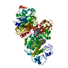
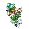
- PDB-4fkz: Crystal structure of Bacillus subtilis UDP-GlcNAc 2-epimerase in ... -
+ Open data

- Basic information
| Entry | Database: PDB / ID: 4fkz | ||||||
|---|---|---|---|---|---|---|---|
| Title | Crystal structure of Bacillus subtilis UDP-GlcNAc 2-epimerase in complex with UDP-GlcNAc and UDP | ||||||
 Components Components | UDP-N-acetylglucosamine 2-epimerase | ||||||
 Keywords Keywords | BIOSYNTHETIC PROTEIN | ||||||
| Function / homology |  Function and homology information Function and homology informationUDP-N-acetylglucosamine 2-epimerase (non-hydrolysing) / UDP-N-acetylglucosamine 2-epimerase activity / teichoic acid biosynthetic process / polysaccharide biosynthetic process / cell wall organization / cytosol Similarity search - Function | ||||||
| Biological species |  | ||||||
| Method |  X-RAY DIFFRACTION / SYNCHROTRON / MOLECULAR REPLACEMENT / Resolution: 1.69 Å X-RAY DIFFRACTION / SYNCHROTRON / MOLECULAR REPLACEMENT / Resolution: 1.69 Å | ||||||
 Authors Authors | Yang, C.S. / Chen, S.C. / Kuan, S.M. / Chen, Y.R. / Liu, Y.H. / Chen, Y. | ||||||
 Citation Citation | Journal: To be Published Title: Crystal structure of Bacillus subtilis UDP-GlcNAc 2-epimerase in complex with UDP-GlcNAc and UDP Authors: Yang, C.S. / Chen, S.C. / Kuan, S.M. / Chen, Y.R. / Liu, Y.H. / Chen, Y. | ||||||
| History |
|
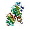
- Structure visualization
| Structure viewer | Molecule: MolmilJmol/JSmol |
|---|
- Downloads & links
-Download
| PDBx/mmCIF format | 4fkz.cif.gz | 186.3 KB | Display | PDBx/mmCIF format |
|---|---|---|---|---|
| PDB format | pdb4fkz.ent.gz | 145.7 KB | Display | PDB format |
| PDBx/mmJSON format | 4fkz.json.gz | Tree view | PDBx/mmJSON format | |
| Others |  Other downloads Other downloads |
-Validation report
| Arichive directory | https://data.pdbj.org/pub/pdb/validation_reports/fk/4fkzftp://data.pdbj.org/pub/pdb/validation_reports/fk/4fkz | HTTPS FTP |
|---|
-Related structure data
| Related structure data | 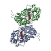 3beoS S: Starting model for refinement |
|---|---|
| Similar structure data |


-Links
 PDBj
PDBj


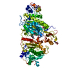
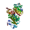
- Assembly
| Deposited unit | 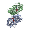
| ||||||||
|---|---|---|---|---|---|---|---|---|---|
| 1 |
| ||||||||
| Unit cell |
|
-Components
| #1: Protein | Mass: 43764.523 Da / Num. of mol.: 2 Source method: isolated from a genetically manipulated source Source: (gene. exp.) Strain: 168 / Gene: BSU35660, mnaA / Plasmid: pET24a / Production host: References: UniProt: P39131, UDP-N-acetylglucosamine 2-epimerase (non-hydrolysing) #2: Chemical |   Mass: 607.354 Da / Num. of mol.: 2 / Source method: obtained synthetically / Formula: C17H27N3O17P2 Mass: 607.354 Da / Num. of mol.: 2 / Source method: obtained synthetically / Formula: C17H27N3O17P2#3: Chemical |   Type: RNA linking / Mass: 404.161 Da / Num. of mol.: 2 / Source method: obtained synthetically / Formula: C9H14N2O12P2 / Comment: UDP*YM Type: RNA linking / Mass: 404.161 Da / Num. of mol.: 2 / Source method: obtained synthetically / Formula: C9H14N2O12P2 / Comment: UDP*YM#4: Water | ChemComp-HOH / |  Mass: 18.015 Da / Num. of mol.: 1008 / Source method: isolated from a natural source / Formula: H2O Mass: 18.015 Da / Num. of mol.: 1008 / Source method: isolated from a natural source / Formula: H2O |
|---|
-Experimental details
-Experiment
| Experiment | Method: X-RAY DIFFRACTION / Number of used crystals: 1 |
|---|
- Sample preparation
| Crystal | Density Matthews: 2.55 Å3/Da / Density % sol: 51.86 % |
|---|---|
| Crystal grow | Temperature: 277 K / Method: vapor diffusion, sitting drop / pH: 7.5 Details: 70% MPD, 0.1 M HEPES, pH 7.5, VAPOR DIFFUSION, SITTING DROP, temperature 277K |
-Data collection
| Diffraction | Mean temperature: 100 K |
|---|---|
| Diffraction source | Source: SYNCHROTRON / Site: NSRRC  / Beamline: BL13C1 / Wavelength: 0.97622 Å / Beamline: BL13C1 / Wavelength: 0.97622 Å |
| Detector | Type: ADSC QUANTUM 210 / Detector: CCD / Date: Dec 3, 2011 |
| Radiation | Monochromator: Si(111) / Protocol: SINGLE WAVELENGTH / Monochromatic (M) / Laue (L): M / Scattering type: x-ray |
| Radiation wavelength | Wavelength: 0.97622 Å / Relative weight: 1 |
| Reflection | Resolution: 1.69→50 Å / Num. all: 101913 / Num. obs: 99875 / % possible obs: 98 % / Redundancy: 3.6 % / Rmerge(I) obs: 0.057 / Net I/σ(I): 18.53 |
| Reflection shell | Resolution: 1.69→1.75 Å / Redundancy: 3.4 % / Rmerge(I) obs: 0.426 / Mean I/σ(I) obs: 3.07 / Num. unique all: 9869 / % possible all: 98.2 |
- Processing
| Software |
| |||||||||||||||||||||||||||||||||||||||||||||
|---|---|---|---|---|---|---|---|---|---|---|---|---|---|---|---|---|---|---|---|---|---|---|---|---|---|---|---|---|---|---|---|---|---|---|---|---|---|---|---|---|---|---|---|---|---|---|
| Refinement | Method to determine structure: MOLECULAR REPLACEMENT Starting model: PDB ENTRY 3BEO Resolution: 1.69→32.29 Å / Cor.coef. Fo:Fc: 0.966 / Cor.coef. Fo:Fc free: 0.954 / SU B: 1.776 / SU ML: 0.06 / Cross valid method: THROUGHOUT / σ(F): 0 / ESU R: 0.097 / ESU R Free: 0.094 / Stereochemistry target values: MAXIMUM LIKELIHOOD Details: HYDROGENS HAVE BEEN USED IF PRESENT IN THE INPUT U VALUES : REFINED INDIVIDUALLY
| |||||||||||||||||||||||||||||||||||||||||||||
| Solvent computation | Ion probe radii: 0.8 Å / Shrinkage radii: 0.8 Å / VDW probe radii: 1.2 Å / Solvent model: MASK | |||||||||||||||||||||||||||||||||||||||||||||
| Displacement parameters | Biso mean: 21.704 Å2
| |||||||||||||||||||||||||||||||||||||||||||||
| Refinement step | Cycle: LAST / Resolution: 1.69→32.29 Å
| |||||||||||||||||||||||||||||||||||||||||||||
| Refine LS restraints |
| |||||||||||||||||||||||||||||||||||||||||||||
| LS refinement shell | Resolution: 1.69→1.732 Å / Total num. of bins used: 20
|
