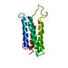
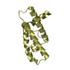
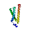
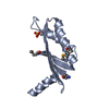
Entry Database : PDB / ID : 4f2lTitle Structure of a regulatory domain of AMPK 5'-AMP-activated protein kinase catalytic subunit alpha-1 Keywords / / / Function / homology Function Domain/homology Component
/ / / / / / / / / / / / / / / / / / / / / / / / / / / / / / / / / / / / / / / / / / / / / / / / / / / / / / / / / / / / / / / / / / / / / / / / / / / / / / / / / / / / / / / / / / / / / / / / / / / / / / / / / / / / / / / / / / / / / / / / / / / / / / / / / / / / / / / / Biological species Rattus norvegicus (Norway rat)Method / / Resolution : 1.5 Å Authors Xin, F.J. / Zhang, Y.Y. / Wang, J. / Wang, Z.X. / Wu, J.W. Journal : Nature / Year : 2013Title : Conserved regulatory elements in AMPKAuthors : Chen, L. / Xin, F.J. / Wang, J. / Hu, J. / Zhang, Y.Y. / Wan, S. / Cao, L.S. / Lu, C. / Li, P. / Yan, S.F. / Neumann, D. / Schlattner, U. / Xia, B. / Wang, Z.X. / Wu, J.W. History Deposition May 8, 2012 Deposition site / Processing site Revision 1.0 Jun 26, 2013 Provider / Type Revision 1.1 Sep 3, 2014 Group Revision 1.2 Nov 15, 2017 Group / Category Revision 1.3 Oct 16, 2024 Group Data collection / Database references ... Data collection / Database references / Derived calculations / Structure summary Category chem_comp_atom / chem_comp_bond ... chem_comp_atom / chem_comp_bond / database_2 / pdbx_entry_details / pdbx_modification_feature / pdbx_struct_conn_angle / struct_conn / struct_ref_seq_dif / struct_site Item _database_2.pdbx_DOI / _database_2.pdbx_database_accession ... _database_2.pdbx_DOI / _database_2.pdbx_database_accession / _pdbx_struct_conn_angle.ptnr1_auth_seq_id / _pdbx_struct_conn_angle.ptnr3_auth_seq_id / _pdbx_struct_conn_angle.value / _struct_conn.pdbx_dist_value / _struct_conn.pdbx_leaving_atom_flag / _struct_conn.ptnr2_auth_seq_id / _struct_ref_seq_dif.details / _struct_site.pdbx_auth_asym_id / _struct_site.pdbx_auth_comp_id / _struct_site.pdbx_auth_seq_id
Show all Show less
 Movie
Movie Controller


 Open data
 Basic information
 Components
 Keywords
 Function and homology information
 X-RAY DIFFRACTION /
  Authors
 Citation
 Structure visualization
 Downloads & links
 Other downloads
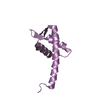


 PDBj
PDBj Assembly
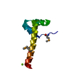


 Mass: 24.305 Da / Num. of mol.: 1 / Source method: obtained synthetically / Formula: Mg
 Mass: 18.015 Da / Num. of mol.: 79 / Source method: isolated from a natural source / Formula: H2O
 Sample preparation
 / Beamline: BL17U / Wavelength: 0.9792 Å
 Processing